
Fig. S1
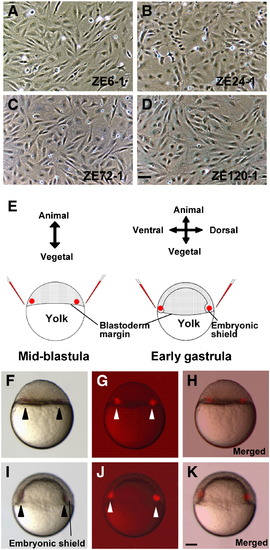
Morphological characteristics of zebrafish cell lines and implantation of these cell lines into mid-blastula or early gastrula. After several passages, each cell line appeared as a fibroblast monolayer. Cells were derived from (A) early gastrula stage embryos (ZE6-1), (B) pharyngula period embryos (ZE24-2), (C) protruding-mouth stage larvae (ZE72-1), and (D) swimming larva period larvae (ZE120-1). (E) Schematic representation of implantation. About 50 DiI-labeled cells per region were implanted into each half of the host marginal region. (F–H) Cell clumps implanted into both halves of the blastoderm margin in the mid-blastula (3 hpf). (I–K) Cell clumps implanted into the shield and the opposite side in early gastrula (6 hpf). (F, I) Bright field image. (G, J) Dark field image. (H, K) Merged images of F and G (H), and I and J (K). All views are lateral. Arrowheads indicate implanted cell clumps. Scale bars are 50 μm (A–D) and 100 μm (F–K). |
Reprinted from Developmental Biology, 321(2), Hashiguchi, M., Shinya, M., Tokumoto, M., and Sakai, N., Nodal/Bozozok-independent induction of the dorsal organizer by zebrafish cell lines, 387-396, Copyright (2008) with permission from Elsevier. Full text @ Dev. Biol.