FIGURE

Fig. 6

ID
ZDB-FIG-080910-19
Publication
Kamei et al., 2008 - Duplication and Diversification of the Hypoxia-Inducible IGFBP-1 Gene in Zebrafish
Other Figures
All Figure Page
Back to All Figure Page
Fig. 6

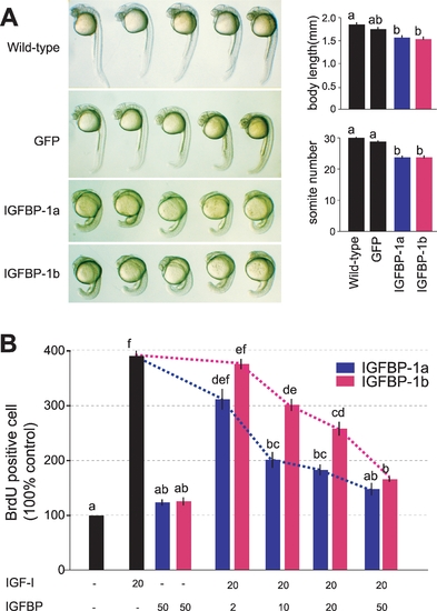
Zebrafish IGFBP-1a and IGFBP-1b are both functional but have different activities.
A) Expression of IGFBP-1a and -1b reduces embryo growth and developmental rate in vivo. GFP mRNA (600 pg/embryo), a mixture of IGFBP-1a mRNA (400 pg/embryo) and GFPmRNA (200 pg/embryo), or IGFBP-1a mRNA (400 pg/embryo) and GFP mRNA (200 pg/embryo) was injected into 1–2 cell stage embryos. The embryos were raised to 24 hpf and body length and somite number were determined. Representative embryos are shown in the left column. Body length and somite number of each experimental group are shown in the left column. Values are represented as means±S.E. (n = 21–29). Groups with common letters are not significantly different from each other (p<0.05). B) Effects of IGFBP-1a and -1b on IGF-I-stimulated cell proliferation in cultured zebrafish cells. Serum starved confluent cells were exposed to the media with or without 20 nM IGF-I in the absence or presence of various concentrations of IGFBP-1s (2, 10, 20 or 50 nM). To evaluate IGF-independent effect, cells are exposed with 50 nM of IGFBP-1s without IGF. The percentage of BrdU-positive cells in each group was calculated and is shown. Values are means±S.E. (n = 3∼6). Groups with common letters are not significantly different from each other (p<0.05).

Expression Data

Expression Detail
Antibody Labeling
Phenotype Data

Phenotype Detail
Acknowledgments
This image is the copyrighted work of the attributed author or publisher, and ZFIN has permission only to display this image to its users. Additional permissions should be obtained from the applicable author or publisher of the image. Full text @ PLoS One