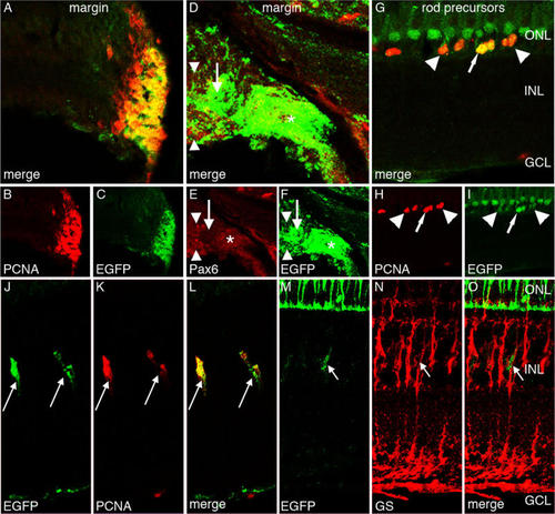
Enhanced green fluorescent protein expression in the Tg(ccnb1:EGFP)nt18 adult zebrafish retina. Enhanced green fluorescent protein (EGFP; C, F, I, J, M), proliferating cell nuclear antigen (PCNA; B, H, K), Pax6 (E), glutamine synthetase (GS; N), and merged expression (A, D, G, L, O) are shown. EGFP and PCNA are strongly expressed in the cells of the adult retinal circumferential marginal zone (CMZ; A-C). EGFP expression in the peripheral CMZ co-labels with Pax6 (*), but does not co-label near the distal CMZ (D-F, arrow). The Pax6-positive EGFP-negative signal adjacent to the CMZ (arrowheads) corresponds to the newly differentiated amacrine and ganglion cells. EGFP is also expressed in PCNA-positive rod precursor cells, which reside in the outer nuclear layer (G-I). Some of these rod precursor cells express high levels of EGFP (arrows), while others express lower EGFP levels (arrowhead). EGFP is also expressed in a row of nonproliferative PCNA-negative short single cones (G, I). Some Müller glia in the adult undamaged retina slowly divide and give rise to retinal progenitor cells. PCNA marks these proliferating Müller glial cells, which also coexpress EGFP (J-L, arrows). EGFP-positive cells in the INL also co-label with glutamine synthetase (M-O, arrow). This demonstrates that proliferating EGFP-positive cells co-label with Müller glia. Abbreviations: ONL represents outer nuclear layer; INL represents inner nuclear layer; and GCL represents ganglion cell layer.
|
