
Fig. 4
- ID
- ZDB-FIG-080424-62
- Publication
- Dorsky et al., 2002 - A transgenic Lef1/beta-catenin-dependent reporter is expressed in spatially restricted domains throughout zebrafish development
- Other Figures
- All Figure Page
- Back to All Figure Page
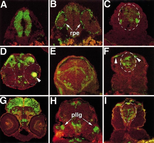
Transverse cryosections of transgenic embryos show localized expression in regions of the CNS and other tissues. In all panels, red autofluorescence is shown for contrast. Spinal cord is outlined in white dotted lines in (C, F, I). (A–C) 24 hpf; (D–F) 36 hpf; (G–I) 48 hpf. (A) Midbrain rostral to the eye is shown. (B) Section through caudal eye region. rpe, retinal pigmented epithelium. (C) Spinal cord section, showing individual GFP-labeled neurons. (D) Eye and midbrain region. Eyes are outlined in white dotted lines, lens is indicated by arrowhead. (E) Hindbrain section. Individual neurons in the ventral hindbrain express GFP. (F) In the spinal cord, expression is seen at multiple dorsal/ventral positions and in dorsal pigment cells (arrowheads). Pigment cells are identifiable by their morphology and position just below the ectoderm. (G) Extensive GFP expression is present in the dorsal midbrain. (H) Section just caudal to ear. pllg, posterior lateral line ganglion. (I) Spinal cord expression is similar to domains observed at 36 hpf. |
Reprinted from Developmental Biology, 241(2), Dorsky, R.I., Sheldahl, L.C., and Moon, R.T., A transgenic Lef1/beta-catenin-dependent reporter is expressed in spatially restricted domains throughout zebrafish development, 229-237, Copyright (2002) with permission from Elsevier. Full text @ Dev. Biol.