Fig. 2
- ID
- ZDB-FIG-080424-57
- Publication
- Hsu et al., 2002 - Expression of zebrafish cyp11a1 as a maternal transcript and in yolk syncytial layer
- Other Figures
- All Figure Page
- Back to All Figure Page
|
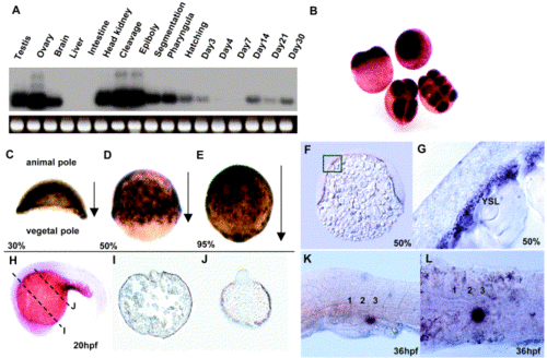
Expression of zebrafish cyp11a1 during embryogenesis. (A) RT–PCR analysis followed by hybridization of cyp11a1 in adult tissues and at various developmental stages: cleavage (1 to 128-cell), epiboly (sphere, 30, 50, and 70% epiboly), segmentation (12 and 18 hpf), pharyngula (24 hpf), hatching (48 hpf). RT–PCR of actin was used as RNA loading control. (B–L) In situ hybridization of cyp11a1: (B) in blastodiscs at the cleavage stage, (C–E) during epiboly below the blastomere extending from the animal pole toward the vegetal pole, (F) sections at 50% epiboly, (G) higher magnification of the square area in (F). (H–J) Expression of cyp11a1 in the yolk syncytial layer (YSL) persists at 20 phf. (I,J) Sections from (H). (K–L) Expression of cyp11a1 is located at a region ventral to the third somite at 36 hpf. Somite numbers are marked as 1, 2, and 3. (K) Lateral view, (L) ventral view. |
Reprinted from Gene expression patterns : GEP, 2(3-4), Hsu, H.J., Hsiao, P., Kuo, M.W., and Chung, B.C., Expression of zebrafish cyp11a1 as a maternal transcript and in yolk syncytial layer, 219-222, Copyright (2002) with permission from Elsevier. Full text @ Gene Expr. Patterns

