
Fig. 4
- ID
- ZDB-FIG-080326-108
- Publication
- Devine et al., 2008 - Robo-Slit interactions regulate longitudinal axon pathfinding in the embryonic vertebrate brain
- Other Figures
- All Figure Page
- Back to All Figure Page
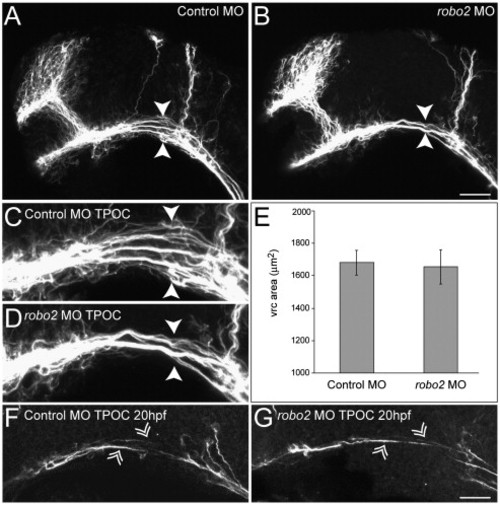
Knockdown of robo2 leads to a partial collapse of the TPOC. Dissected brains are labeled with acetylated α-tubulin at 28 hpf to show all axons. Rostral is to the left and dorsal is to the top. (A) The axon scaffold developed normally in embryos injected with 5 ng of control MO. By contrast, following knockdown of robo2 (5 ng MO2) the TPOC appeared to collapse along its dorsoventral axis (B, arrowheads; compare higher power images in panels C and D). (E) Quantification of the vrc surface area by HuC staining confirmed that the collapsed TPOC phenotype was not associated with a reduction in the size of this neuronal cluster. Values represent the mean ± SD. Significance was assessed using the Student′s t-test. (F) No qualitative differences were observed in the outgrowth of TPOC pioneer axons (double arrowheads) between control MO (5 ng; F) and robo2 MO2 (5 ng, G) scaffolds at 18–20 hpf. Scale bar: A, B: 50 μm; C, D, F, G: 20 μm. |
| Fish: | |
|---|---|
| Knockdown Reagent: | |
| Observed In: | |
| Stage: | Prim-5 |
Reprinted from Developmental Biology, 313(1), Devine, C.A., and Key, B., Robo-Slit interactions regulate longitudinal axon pathfinding in the embryonic vertebrate brain, 371-383, Copyright (2008) with permission from Elsevier. Full text @ Dev. Biol.