
Fig. S3
- ID
- ZDB-FIG-080325-72
- Publication
- Wilkins et al., 2008 - Mtx2 directs zebrafish morphogenetic movements during epiboly by regulating microfilament formation
- Other Figures
- All Figure Page
- Back to All Figure Page
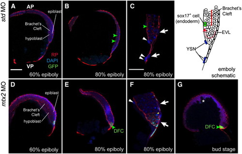
Involution of the blastoderm is not Mtx2-dependent. Vibratome sections of Tg[sox17:EGFP] embryos (A–C) show that Brachet's cleft can be seen from 60% epiboly onwards by rhodamine–phalloidin staining. Involution, shown in the schematic (top right), still occurs in mtx2 MO injected Tg[sox17:EGFP] embryos (D–G), however, the blastoderm is thicker, presumably because the blastoderm does not migrate as far vegetally. Green arrowheads indicate GFP+ cells, white arrowheads indicate YSN, white arrows indicate EVL cells, white asterisk highlights the irregular shape of the blastoderm at the animal pole. DFC = dorsal forerunner cells (not visible in standard controls). Scale bar in A = 150 μm (for A, B, D, E, G). Scale bar in C = 100 μm (for C, F). |
| Gene: | |
|---|---|
| Fish: | |
| Knockdown Reagent: | |
| Anatomical Terms: | |
| Stage Range: | 75%-epiboly to Bud |
| Fish: | |
|---|---|
| Knockdown Reagent: | |
| Observed In: | |
| Stage Range: | 75%-epiboly to Bud |
Reprinted from Developmental Biology, 314(1), Wilkins, S.J., Yoong, S., Verkade, H., Mizoguchi, T., Plowman, S.J., Hancock, J.F., Kikuchi, Y., Heath, J.K., and Perkins, A.C., Mtx2 directs zebrafish morphogenetic movements during epiboly by regulating microfilament formation, 12-22, Copyright (2008) with permission from Elsevier. Full text @ Dev. Biol.