
Fig. 3
- ID
- ZDB-FIG-071204-22
- Publication
- Yokoi et al., 2007 - Mutant analyses reveal different functions of fgfr1 in medaka and zebrafish despite conserved ligand-receptor relationships
- Other Figures
- All Figure Page
- Back to All Figure Page
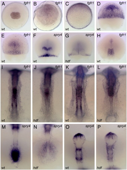
Embryonic expression of medaka fgfr1 and its down-stream target, sprouty4. Probes used are on the top right corner. wt, wild-type and hdf, fgfr1hdf mutant embryos. Animal-pole views (A–B) and dorsal views (E–P; animal-pole or anterior to the top) are shown. Maternal expression of medaka fgfr1 is detected ubiquitously at st. 4 (4-cell) (A). At st. 13 (early gastrula), fgfr1 is expressed ubiquitously with higher levels on the dorsal side (to the top; C). During gastrulation, fgfr1 is expressed in the presumptive head region with the continued expression in the margin (D, st. 15; E, st. 15+). The expression of sprouty4 is observed in the presumptive MHB and dorsal margin in a part of the sibling embryos (F), but in some siblings this expression is undetectable (G). At the end of gastrulation, fgfr1 is expressed in the trunk region (H). At the bud stage (I) and the early segmentation stage (K), fgfr1 is expressed in the trunk, paraxial mesoderm, somite and presomitic mesoderm. In the neural tube, weak ubiquitous expression with high levels in the hindbrain is observed in the wild-type embryo (I, K). In the mutant, fgfr1 expression is lost in the paraxial mesoderm, somites and presomitic mesoderm, due to a lack of these tissues, whereas the expression in the anterior portion remains unchanged (J, L). At these stages, sprouty4 is expressed in the tailbud (M), the anterior tip of telencephalon, MHB and r4 (O). The sprouty4 expression is lost in the tailbud and reduced in the mutant (N, P), except for the telencephalon. |
Reprinted from Developmental Biology, 304(1), Yokoi, H., Shimada, A., Carl, M., Takashima, S., Kobayashi, D., Narita, T., Jindo, T., Kimura, T., Kitagawa, T., Kage, T., Sawada, A., Naruse, K., Asakawa, S., Shimizu, N., Mitani, H., Shima, A., Tsutsumi, M., Hori, H., Wittbrodt, J., Saga, Y., Ishikawa, Y., Araki, K., and Takeda, H., Mutant analyses reveal different functions of fgfr1 in medaka and zebrafish despite conserved ligand-receptor relationships, 326-337, Copyright (2007) with permission from Elsevier. Full text @ Dev. Biol.