
Fig. 5
- ID
- ZDB-FIG-071019-5
- Publication
- Fan et al., 2007 - Nodal signals mediate interactions between the extra-embryonic and embryonic tissues in zebrafish
- Other Figures
- All Figure Page
- Back to All Figure Page
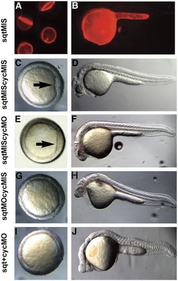
sqt and cyc have partially overlapping roles in the YSL. Images of live 5 hpf (A), 6 hpf (C, E, G, I), or 24 hpf (B, D, F, H) embryos after targeted co-injection into the YSL of sqtMO and cycMO, or appropriate 5 bp mismatched control MOs. After co-injection of sqt and cyc mismatched MOs, the embryonic shield forms normally (A, arrow) and the body axis is indistinguishable from wild type (B). After co-injection of the sqtMIS MO and cycMO, the embryonic shield forms (C, arrow) and the body axis is normal (D). Embryonic shields do not form in many embryos following injection of the sqtMO and cycMIS MO (G). The majority of these embryos appear normal at 24 hpf (H), but many have reduced cardiac tissue. (I, J) After co-injection of sqtMO and cycMO, the vast majority of embryos lack embryonic shields (G). At 24 hpf, these embryos display severe cyclopia and have reduced notochord due to defects in axial mesoderm patterning (J). Paraxial mesoderm is less affected, as somites form in embryos co-injected with sqtMO and cycMO (J). |
| Fish: | |
|---|---|
| Knockdown Reagents: | |
| Observed In: | |
| Stage Range: | Shield to Prim-5 |
Reprinted from Developmental Biology, 310(2), Fan, X., Hagos, E.G., Xu, B., Sias, C., Kawakami, K., Burdine, R.D., and Dougan, S.T., Nodal signals mediate interactions between the extra-embryonic and embryonic tissues in zebrafish, 363-378, Copyright (2007) with permission from Elsevier. Full text @ Dev. Biol.