Fig. 5
- ID
- ZDB-FIG-071001-5
- Publication
- Latimer et al., 2006 - Notch signaling regulates midline cell specification and proliferation in zebrafish
- Other Figures
- All Figure Page
- Back to All Figure Page
|
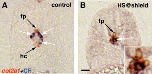
Notch signaling drives trunk axial mesoderm to a hypochord cell fate. (A, B) 24 hpf, transverse sections, showing col2a1 expression combined with staining to show photoactivated fluorescein signal. Three to five cells were labeled by photoactivation of caged fluorescein at the dorsal margin in control embryos and embryos expressing NICD beginning just before 6 hpf. (A) Labeled cells were found in the notochord (white arrow) and never in col2a1-expressing hypochord. (B) Cells derived from the notochord precursor domain expressed col2a1 following heat shock activation of Notch at 6 hpf. Inset shows enlarged view of midline region. Scale bar: 10 μm for panels A, B. |
Reprinted from Developmental Biology, 298(2), Latimer, A.J., and Appel, B., Notch signaling regulates midline cell specification and proliferation in zebrafish, 392-402, Copyright (2006) with permission from Elsevier. Full text @ Dev. Biol.

