
Fig. 4
- ID
- ZDB-FIG-070925-24
- Publication
- Yang et al., 2007 - A highly conserved regulatory element controls hematopoietic expression of GATA-2 in zebrafish
- Other Figures
- All Figure Page
- Back to All Figure Page
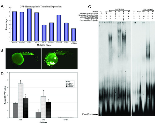
Conservation of Up2 enhancer activity and transacting factors. (A). Transient transgenic assays of hematopoietic GFP expression in embryos injected with wild type and mutant constructs identify HOXA3, LMO2 and E2F-1 as potential transcription factors. Also see Additional file 2, Supplementary Table S for more information about the results of hematopoietic transient expression assay. (B). Typical transient GFP expression patterns in injected embryos at 22 hpf. Left, GFP expression is detected in hematopoietic cells (arrow); Right, non-specific GFP expression was observed in muscle, heart and neuron tissue but not in hematopoietic cells (C). EMSA shows transcription factor E2F-1 (left) and HOXA3 (right) specifically bind to the probe corresponding to Up2 sequence, respectively. Probe containing HOXA3-A binding site was analyzed in EMSA. (D). Human myeloid leukemia cell lines, KG1 and K562, and mouse fibroblast cell line, NIH3T3 were transiently transfected with the mp/GFP, Up2-mp/GFP and Up2a-mp/GFP constructs. GFP expression was stronger in myeloid cells than in the fibroblast cell line. In the myeloid cell lines, enhancer activity of Up2 and Up2a was found to be statistically significant (*p < 0.002, ^p < 0.05, # p < 0.035, ** p < 0.032). The enhancer activity of Up2a/mp was less than that of Up2/mp. This figure represents the average of three independent experiments performed in duplicate. |