
Fig. 1
- ID
- ZDB-FIG-070911-11
- Publication
- Malone et al., 2007 - Laser-scanning velocimetry: A confocal microscopy method for quantitative measurement of cardiovascular performance in zebrafish embryos and larvae
- Other Figures
- All Figure Page
- Back to All Figure Page
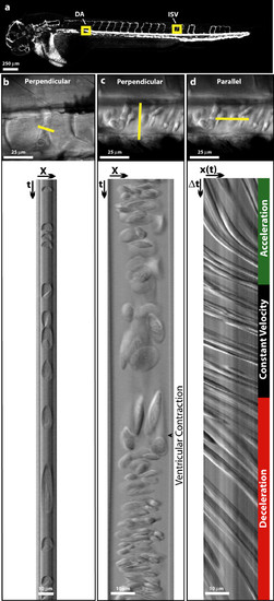
Acquring linescan images from Danio rerio embryos. (a) In silico microangiogram of a 48 hpf wild-type zebrafish embryo. Regions of interest used for acquiring linescan images are boxed (intersomitic vessel = ISV, dorsal aorta = DA). (b and c) A single plane (XY) image (Top) showing the orientation of scan lines used to create perpendicular linescans (Bottom) of the intersomitic vessels (b) and dorsal aorta (c). The boundary between slow moving (elongated) cells and fast moving (compressed) cells reflects a ventricular contraction and is easily observed in the Dorsal Aorta (c). (d) A parallel linescan acquired from the dorsal aorta shows a series of lines whose slope is inversely proportional to cellular velocity. Distinct regions of acceleration, constant velocity, and deceleration are observed. |