
Fig. 3
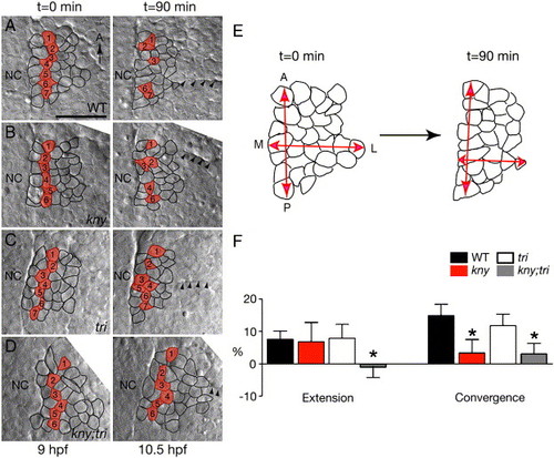
C&E movements of the prospective adaxial cell population during late gastrulation. (A–D) Nomarski images of live embryos captured at the beginning and the end of the time-lapse recordings. Dorsal views, anterior to the top. Cells located two-cell diameters away from the notochord at the beginning of the time-lapse are labeled in red. Their positions at the end of the time-lapses are indicated in the right panels. Arrowheads point to the first somitic boundary. (E) Schematic of the tissue shape change in WT. (F) Quantifications of the tissue shape changes (Materials and methods). Error bars represent the standard error. Asterisk: p < 0.05, mutant versus WT. NC, notochord. M, medial. L, lateral. Scale bar: 50 μm. |
Reprinted from Developmental Biology, 304(1), Yin, C., and Solnica-Krezel, L., Convergence and extension movements mediate the specification and fate maintenance of zebrafish slow muscle precursors, 141-155, Copyright (2007) with permission from Elsevier. Full text @ Dev. Biol.