
Fig. 2
- ID
- ZDB-FIG-070802-41
- Publication
- Molina et al., 2007 - Generation of FGF reporter transgenic zebrafish and their utility in chemical screens
- Other Figures
- All Figure Page
- Back to All Figure Page
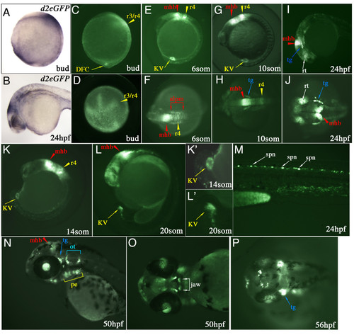
Spatial and temporal d2EGFP expression in Tg(Dusp6:d2EGFP)pt6 embryos. (A & B) Lateral views of d2EGFP mRNA expression at Bud stage and 24 hpf. (C-P) d2EGFP expression in Tg(Dusp6:d2EGFP)pt6 embryos, stages are indicated in each panel. At bud stage (C & D), d2EGFP is detected in the hindbrain (r3/r4, yellow arrowhead) and within the caudal region in the DFCs. (E, G & K) From 8- to 14-somite stages, lateral views show expression of d2EGFP in cells lining Kupffer's vesicle, within r4 (r4, yellow arrowhead) and the mid-hindbrain boundary (mhb, red arrowhead). (F & H) Dorsal views show high d2EGFP expression within the MHB, r4 and the anterior lateral plate mesoderm (alpm, red brackets). (H) At 10-somite stage initial d2EGFP expression is detected within the trigeminal ganglia (tg, blue arrow). (I & J) 24 hpf embryo showing d2EGFP expression in the MHB, trigeminal ganglia, dorsal retina (rt, white arrow) and pharyngeal endoderm (pe, yellow bracket). (K & L) 14 and 20-somite stage embryo highlighting the expression of d2EGFP in Kupffer's vesicle. Higher magnifications are show in (K' & L'). (M) Trunk region shows d2EGFP expression within the dorsal spinal cord neurons (spn, white arrow) at 24 hpf. (N) At 50 hpf expression is noted in the MHB, trigeminal ganglia, pharyngeal endoderm and otic vesicle (ot, blue bracket). (O) Ventral view of 50 hpf, showing d2EGFP expression in the jaw (white bracket). (P) At 56 hpf, strong expression in noted in the trigeminal ganglia, the jaw and also in neurons within the dorsal diencephalon. |