
Fig. 3
- ID
- ZDB-FIG-070613-34
- Publication
- Kawaguchi et al., 2007 - Analysis of the exon-intron structures of fish, amphibian, bird and mammalian hatching enzyme genes, with special reference to the intron loss evolution of hatching enzyme genes in Teleostei
- Other Figures
- All Figure Page
- Back to All Figure Page
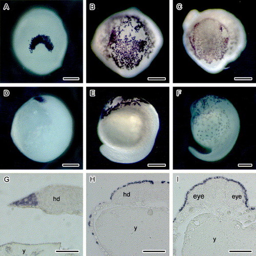
Whole-mount in situ hybridization: The expression of FgHCE and FgLCE gene during the development of fugu embryos. FgHCE (A–C and E) and FgLCE (D and F) RNA probes were hybridized with 3 somite embryos (A and D), 12–15 somite embryos (B and E) and 22–23 somite embryos (C and F). A–C: views of head regions. Upper, the anterior-most. D–F: lateral views. Right, dorsal. The sections of embryos hybridized with the FgHCE (H–I) and FgLCE probe (G) were made from the 3 somite embryos (G) and 12–15 somite embryos (H and I). G–H: sagittal section, I: transverse section. Scale bars, A–F; 200 μm; G–I; 100 μm. y, yolk; hd, head. |
Reprinted from Gene, 392(1-2), Kawaguchi, M., Yasumasu, S., Hiroi, J., Naruse, K., Suzuki, T., and Iuchi, I., Analysis of the exon-intron structures of fish, amphibian, bird and mammalian hatching enzyme genes, with special reference to the intron loss evolution of hatching enzyme genes in Teleostei, 77-88, Copyright (2007) with permission from Elsevier. Full text @ Gene