
Fig. 1
- ID
- ZDB-FIG-061221-1
- Publication
- Wagner et al., 2002 - Modulation of BMP activity in dorsal-ventral pattern formation by the Chordin and Ogon antagonists
- Other Figures
- All Figure Page
- Back to All Figure Page
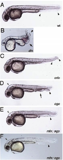
mini fin (mfn) double mutant analysis. Phenotypes observed in embryos from crosses between mfn;din double heterozygous fish. (A) In wild type embryos a single ventral tail fin extends the full length of the tail (arrowheads). (B) The din mutant displays multiple ventral tail fins (arrowhead), an edema posterior to the yolk extension at the site of blood formation (arrow) and a variably reduced head and eyes. (C) mfn mutant embryos display a gap in the ventral tail fin (arrowheads). Additional phenotypes to (A and C) observed in embryos from crosses of mfn;ogon double heterozygous fish (D and E). (D) The ogon mutant is characterized by multiple ventral tail fins (black arrowhead), an edema posterior to the yolk extension (arrow) and an altered head morphology (white arrowhead). (E) Embryos exhibiting the ogon head phenotype (white arrowhead) and a much less severe or wild type tail phenotype (black arrow head). This embryo had only a small edema posterior to the yolk extension without the pooling of blood or the multiplicated ventral tail fins seen in D. |
Reprinted from Developmental Biology, 245(1), Wagner, D.S., and Mullins, M.C., Modulation of BMP activity in dorsal-ventral pattern formation by the Chordin and Ogon antagonists, 109-123, Copyright (2002) with permission from Elsevier. Full text @ Dev. Biol.