FIGURE

Fig. 2

ID
ZDB-FIG-061104-17
Publication
Sakaguchi et al., 2006 - The yolk syncytial layer regulates myocardial migration by influencing extracellular matrix assembly in zebrafish
Other Figures
All Figure Page
Back to All Figure Page
Fig. 2

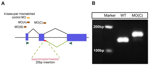
Specificity of mtx1 MOs. (A) Schematic diagram of mtx1 genomic structure and position of mtx1 MOs. mtx1 has three exons (blue boxes) and two introns. Two independent ATG MOs, MO(A) and MO(B), and a splice donor MO, MO(C), caused essentially the same phenotype when injected into the YSL at the 1000-cell stage. The splice donor MO, mtx1 MO(C), leads to a 20 bp insertion in mtx1 mRNA, which leads to a frame shift and premature termination. Green arrowheads indicate the position of the primers used for the RT-PCR analysis shown in B. (B) RT-PCR analysis shows that an additional 20 bp are integrated into mtx1 mRNA in mtx1 MO(C)-injected embryos.

Expression Data
Gene:
Fish:
Knockdown Reagent:
Anatomical Term:
Stage: Shield

Expression Detail
Antibody Labeling
Phenotype Data

Phenotype Detail
Acknowledgments
This image is the copyrighted work of the attributed author or publisher, and ZFIN has permission only to display this image to its users. Additional permissions should be obtained from the applicable author or publisher of the image. Full text @ Development