Fig. 6
- ID
- ZDB-FIG-060628-29
- Publication
- Wei et al., 2004 - The zebrafish Pard3 ortholog is required for separation of the eye fields and retinal lamination
- Other Figures
- All Figure Page
- Back to All Figure Page
|
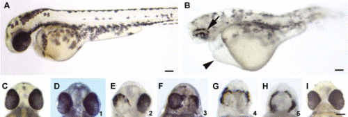
Morpholino-mediated reduction of Pard3 expression caused patchy RPE pigmentation and cyclopia. A wild-type and anti-pard3 morpholino-injected embryo at 2 dpf (A and B, respectively). The patchy pigmentation in the RPE (arrow) and the pericardial edema (arrowhead) in the morpholino-treated embryo are marked. The ventral head region of a wild-type embryo (C) shows the normal spacing between the eyes. A spectrum of cyclopic phenotypes (D–H) in anti-pard3 morpholino-treated embryos exist from the wild-type distance between the eyes (D) to a single eye (H). The numbers in the lower right corner of Panels D–H (ranging from 1 to 5, respectively) represent the degrees of cyclopia expressivity that were used to determine the cyclopia index in Table 2 and Table 3. Co-injection of in vitro-transcribed pard3 mRNA (encoding the 150-kDa Pard3 isoform flanked by the β-globin 5′ and 3′ UTRs) with the anti-pard3 morpholino (Morph-1) rescued the Morph-1-induced cyclopia phenotype and the patchy RPE pigmentation defect (I). Scale bars indicate 100 μm. |
| Fish: | |
|---|---|
| Knockdown Reagent: | |
| Observed In: | |
| Stage: | Long-pec |
Reprinted from Developmental Biology, 269(1), Wei, X., Cheng, Y., Luo, Y., Shi, X., Nelson, S., and Hyde, D.R., The zebrafish Pard3 ortholog is required for separation of the eye fields and retinal lamination, 286-301, Copyright (2004) with permission from Elsevier. Full text @ Dev. Biol.

