
Fig. 3
- ID
- ZDB-FIG-060301-24
- Publication
- Sato-Maeda et al., 2006 - Sema3a1 guides spinal motor axons in a cell- and stage-specific manner in zebrafish
- Other Figures
- All Figure Page
- Back to All Figure Page
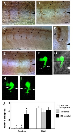
Extension by CaP axons is aberrant following knockdown of Sema3a1. (A) CaP axons extend normally in a control sema3a1 MO-injected embryo (24 hpf) seen in a lateral view. Arrowhead indicates the nascent horizontal myoseptum and broken lines indicate somitic segmental borders. (B) CaP axons often branch aberrantly in an antisense sema3a1 MO-injected embryo (24 hpf). (C) CaP axons can stall (asterisk) in an antisense sema3a1 MO-injected embryo (24 hpf). (D) Transverse section showing a CaP axon extending abnormally into the lateral myotome in the horizontal myoseptal region (arrow) in an antisense sema3a1 MO-injected embryo (24 hpf). Sc, spinal cord; Nc, notochord. (E) Aberrantly branched CaP axons in an antisense nrp1a MO-injected embryo (24 hpf). (F) GFP-labeled CaP neurons in a nrp1a:gfp transgenic embryo showing filopodia from the leading edge of the growth cone (arrows) but not the axon behind the growth cone. (G) Nomarski bright-field image is overlaid to show the location of the CaP growth cone. White arrowhead indicates the horizontal myoseptum. (H,I) Two independent examples showing the same feature. Filopodia extending from the growth cone and trailing axon (arrows) of CaP neurons in a nrp1a:gfp transgenic embryo following injection of antisense sema3a1 MO. (J) Histogram showing an increase in lateral filopodia in CaP axons in the proximal half of the axon in Sema3a1 morphant embryos compared with uninjected embryos and control morphants. Filopodia were quantified from 10 µm CaP axons that were divided into proximal and distal halves. Bars indicate s.d. *P<0.002 for Student's t-test between CaP axons in antisense sema3a1 morphants (n=6) versus control morphants (n=5) and uninjected embryos (n=6). Scale bars: 20 µm. |