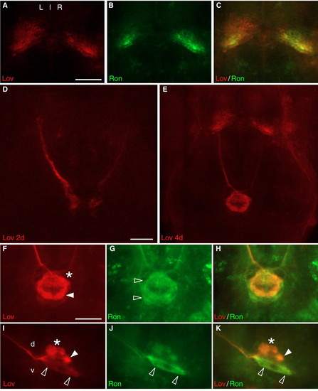
Asymmetric distribution of Lov and Ron in habenular neurons and their projections. (A-C) Immunofluorescent detection of (A) Lov and (B) Ron proteins and (C) colocalization in the habenular nuclei correlates well with the pattern of transcriptional domains (see Fig. 1). (D) Growth cones of Lov+ habenular efferents in FR reach the ventral midbrain by 2 days. (E) By 4 days, asymmetric Lov+ habenular projections traverse the target IPN. (F,I) Lov+ and (G,J) Ron+ axons project to different domains along dorsoventral axis of IPN. (I) Lov+ neurons terminate in anterior (asterisk) and posterior (closed arrowhead) regions of dorsal IPN and in ventral IPN (open arrowheads); (J) most Ron+ neurons project to ventral IPN. (Ron+ immunofluorescence at dorsal IPN was barely distinguishable from background levels, which were always higher than observed with the Lov antiserum.) All images are confocal Z-stacks from (A-H) the dorsal aspect, anterior to top, or (I-K) the lateral aspect, anterior to left. Scale bars: 30 µm (A-C,F-K); 50 µm (D,E).
|
