FIGURE SUMMARY
Title

Apigenin promotes melanogenesis and melanosome transport through the c-KIT/Raf-1/MAPK/CREB pathway in HEMCs

Authors
Lv, J., Meng, D., Zhang, H., Xu, W., An, X., Yin, C., Zou, K., Gao, R.
Source

Apigenin induces hyperpigmentation in melanocytes. (A) Chemical structure of apigenin. (B) HEMCs were treated with various concentrations (0–20 μM) of apigenin for 48 h, and cell viability was assessed using the MTT assay. (C) HEMCs were treated with apigenin (0, 1, 5, or 10 μM) for 48 h, and melanin content was measured. (D) HEMCs were treated with apigenin (10 μM) or α-MSH (100 nM, positive control) for 48 h, followed by Masson-Fontana ammoniacal silver staining. Scale bar = 10 μm. The total dendrite length per cell was measured, and twenty cells per condition were analyzed in three independent experiments. (E) HEMCs were observed using TEM after 48 h of treatment with apigenin (10 μM). White and black arrows indicate stages I–II and III–IV melanosomes, respectively. The percentages of stages I–II and III–IV melanosomes relative to the total number of melanosomes are shown. Data are expressed as the mean ± SEM (n = 3). *p < 0.05, **p < 0.01, and ***p < 0.001 vs. untreated cells. HEMCs, human epidermal melanocytes; α-MSH, α-melanocyte-stimulating hormone; TEM, Transmission electron microscopy.

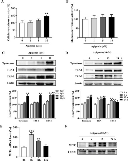
Apigenin increases cellular tyrosinase activity and expression of melanogenic proteins. (A) Cellular tyrosinase activity was measured after treatment with apigenin (0, 1, 5, or 10 μM). (B) Mushroom tyrosinase activity was measured in a cell-free assay after treatment with apigenin (0, 1, 5, or 10 μM). (C) HEMCs were treated with apigenin (0, 1, 5, or 10 μM) for 48 h, and Western blotting was performed to assess protein levels of melanogenic markers. (D) Time-dependent changes in the expression levels of various proteins after treatment with apigenin (10 μM) for the indicated periods. (E) HEMCs were treated with apigenin (10 μM) for 4 h, and RT-qPCR was performed to measure MITF gene expression. (F) HEMCs were treated with apigenin (10 μM) for different time points, followed by Western blotting to assess MITF protein levels. Data are expressed as means ± SEM (n = 3). *p < 0.05, **p < 0.01, and ***p < 0.001 vs. untreated cells. HEMCs, human epidermal melanocytes; MITF, Melanocytes inducing transcription factor.

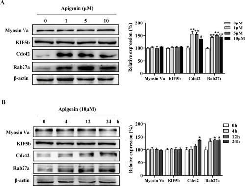
Apigenin promotes the expression of melanosome transport proteins in HEMCs. (A) HEMCs were treated with apigenin (0, 1, 5, or 10 μM) for 48 h, and Western blotting was performed to assess the expression levels of melanosome transport proteins. (B) Time-dependent changes in the expression levels of various melanosome transport proteins following treatment with apigenin (10 μM). Data are expressed as means ± SEM (n = 3). *p < 0.05, **p < 0.01 vs. untreated cells. HEMCs, human epidermal melanocytes.

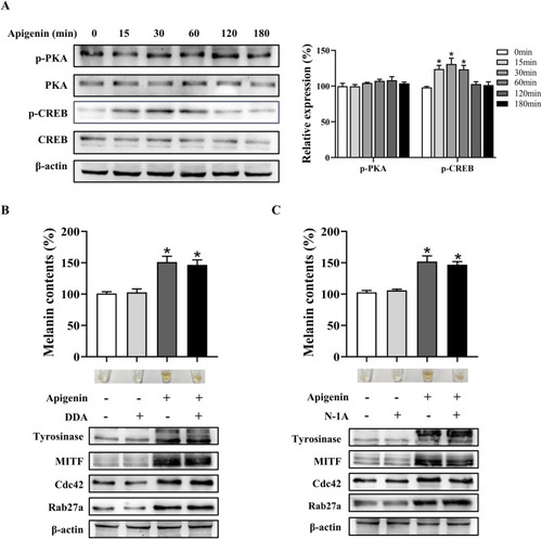
Apigenin promotes pigmentation independent of the classic MC1R/cAMP/PKA-pigmentation pathway in HEMCs. (A) HEMCs were treated with apigenin (10 μM) for the indicated time periods (0–180 min), and the phosphorylation of PKA and CREB was assessed by Western blotting. (B) HEMCs were pretreated with or without 10 μM DDA (an AC inhibitor) for 1 h before the addition of apigenin for 48 h. Melanin content, as well as the expression levels of Tyrosinase, MITF, Cdc42, and Rab27a, were measured as described previously. (C) HEMCs were pretreated with or without 10 μM N-1A (an MC1R inhibitor) for 1 h before apigenin treatment for 48 h. Melanin content and the expression levels of Tyrosinase, Cdc42, and Rab27a were measured as described previously. Data are expressed as the mean ± SEM (n = 3). *p < 0.05 vs. untreated cells. MC1R, melanocortin-1 receptor; cAMP, Cyclic Adenosine Monophosphate; PKA, protein kinase A; HEMCs, human epidermal melanocytes; CREB, cAMP response element-binding protein; AC, adenylate cyclase; MITF, Melanocytes inducing transcription factor; Cdc42, cell division cycle 42; Rab27a, Ras-related protein Rab-27a.

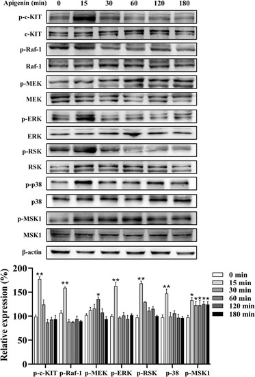
Apigenin activates the c-KIT/Raf-1/MAPK signaling pathway in HEMCs. HEMCs were treated with apigenin (10 μM) for the indicated time periods (0–180 min), and the phosphorylation levels of c-KIT, Raf-1, MEK, ERK, RSK, p38, and MSK1 were assessed by Western blotting. Data are expressed as means ± SEM (n = 3). *p < 0.05, **p < 0.01 vs. untreated cells. c-KIT, cellular-KIT; Raf-1, rapidly accelerated fibrosarcoma-1; MAPK, mitogen-activated protein kinase; HEMCs, human epidermal melanocytes; MEK, mitogen-activated protein kinase kinase; ERK, extracellular signal-regulated kinase; RSK, p90 ribosomal S6 kinase; p38, p38 mitogen-activated protein kinase; MSK1, mitogen-and stress-activated protein kinase 1.

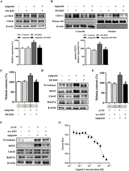
Apigenin promotes pigmentation through the c-KIT-CRTCs/CREB signaling pathway. (A,B) HEMCs were pretreated with or without 10 μM ISCK03 (c-KIT inhibitor) for 1 h before apigenin was added for an additional 1 h. Phosphorylated CREB (p-CREB) and nuclear levels of CRTC1 were measured by Western blotting. Histone H3 served as a reference for nuclear proteins, and β-actin served as a reference for cytosolic proteins. (C,D) HEMCs were pretreated with or without 10 μM ISCK03 for 1 h before apigenin was added for 48 h. Melanin content and the expression levels of Tyrosinase, MITF, Cdc42, and Rab27a were measured as described previously. (E,F) HEMCs were transfected with si-NC or si-c-KIT for 24 h, followed by apigenin (10 μM) treatment for 48 h. Melanin content and the expression levels of Tyrosinase, MITF, Cdc42, and Rab27a were measured as described previously. (G) Binding of c-KIT with apigenin was analyzed by microscale thermophoresis (MST). The binding curve represents data points from 3 measurements. The calculated Kd is 2.6 ± 0.14 µM. Data are expressed as means ± SEM (n = 3). *p < 0.05 vs. non-treated cells, #p < 0.05 vs. apigenin-treated cells. c-KIT, cellular-KIT; CREB, cAMP response element-binding protein; CRTC, CREB-regulated co-activator; HEMCs, human epidermal melanocytes; MITF, Melanocytes inducing transcription factor; Cdc42, cell division cycle 42; Rab27a, Ras-related protein Rab-27a.

Apigenin increases pigmentation in zebrafish. (A) Zebrafish embryos were treated with various concentrations (1–10 μM) of apigenin for different time periods (35–60 h), and zebrafish viability was assessed. (B) Zebrafish embryos were treated with PTU (0.2 mM) for 6–35 h, followed by apigenin (0, 1, 5, or 10 μM) treatment in the embryo medium for 35– 60h. Zebrafish pigmentation was observed using a stereomicroscope. (C) Approximately 30 embryos were collected and dissolved in lysis buffer. After centrifugation, the melanin pigment was dissolved in 200 μL of NaOH working solution (1 mol/L, 10% DMSO) at 80°C for 2 h, and absorbance was measured at 405 nm. Data are expressed as means ± SEM (n = 3). **p < 0.01 vs. non-treated zebrafish, #p < 0.05 and ##p < 0.01 vs. PTU-treated zebrafish. PTU, propylthiouracil; DMSO, Dimethyl Sulfoxide.

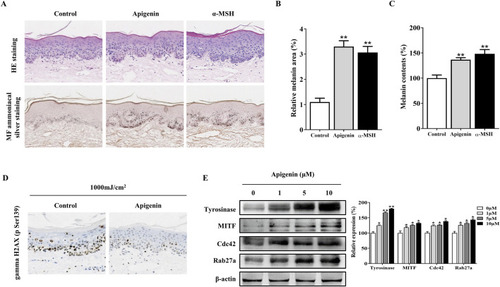
Apigenin induces pigmentation in human skin explants. (A) Human skin explants were treated with apigenin (10 μM) or α-MSH (100 nM) for 5 days. Top panel: H&E staining compared with the vehicle control (DMSO). Bottom panel: Masson-Fontana staining to detect melanin in human skin. (B) The ratio of melanin pigment to the cross-sectional area of human skin, as shown by Masson-Fontana staining. (C) Melanin content was measured in human skin explants treated with apigenin (10 μM) or α-MSH (100 nM) for 5 days. (D) Human skin explants were treated with apigenin for 5 days and exposed to UVB, followed by gamma H2AX (phospho S139) measurement using immunohistochemistry (IHC). (E) Western blotting was performed to assess related protein levels in human skin explants treated with apigenin for 5 days. Data are expressed as the mean ± SEM (n = 3). *p < 0.05, **p < 0.01 vs. non-treated groups. α-MSH, α-melanocyte-stimulating hormone; DMSO, Dimethyl Sulfoxide; UVB, Ultraviolet B.

A proposed model showing that apigenin promotes pigmentation through the c-KIT/Raf-1/MAPK/CREB signaling pathway. Apigenin activates the c-KIT receptor, leading to phosphorylation of Raf-1. Once activated, Raf-1 phosphorylates and activates MAPK, resulting in CREB phosphorylation and subsequent nuclear translocation of CRTCs. Phosphorylated CREB, in association with CRTCs, promotes MITF transcription, which in turn induces the expression of Tyrosinase, Rab27a, and Cdc42. This cascade of events drives melanosome maturation and transport. Tyrosinase, TRP-1, and TRP-2 are key regulators of melanosome maturation. Cdc42 facilitates dendrite extension and filopodia formation, while Rab27a interacts with its effectors, Mlph and Myosin Va, to regulate actin-dependent melanosome transport and anchoring to the plasma membrane. c-KIT, cellular-KIT; Raf-1, rapidly accelerated fibrosarcoma-1; MAPK, mitogen-activated protein kinase; CRTC, CREB-regulated co-activator; CREB, cAMP response element-binding protein; MITF, Melanocytes inducing transcription factor; Cdc42, cell division cycle 42; Rab27a, Ras-related protein Rab-27a; TRP-1, tyrosinase-related protein-1; TRP-2, tyrosinase-related protein-2.

Acknowledgments
This image is the copyrighted work of the attributed author or publisher, and ZFIN has permission only to display this image to its users. Additional permissions should be obtained from the applicable author or publisher of the image.