|
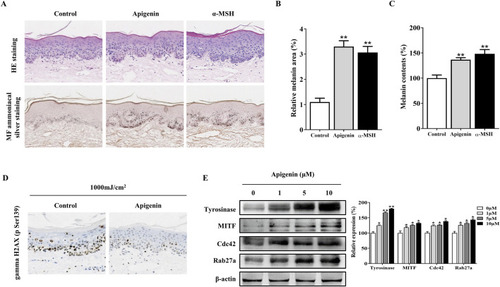
Apigenin induces pigmentation in human skin explants. (A) Human skin explants were treated with apigenin (10 μM) or α-MSH (100 nM) for 5 days. Top panel: H&E staining compared with the vehicle control (DMSO). Bottom panel: Masson-Fontana staining to detect melanin in human skin. (B) The ratio of melanin pigment to the cross-sectional area of human skin, as shown by Masson-Fontana staining. (C) Melanin content was measured in human skin explants treated with apigenin (10 μM) or α-MSH (100 nM) for 5 days. (D) Human skin explants were treated with apigenin for 5 days and exposed to UVB, followed by gamma H2AX (phospho S139) measurement using immunohistochemistry (IHC). (E) Western blotting was performed to assess related protein levels in human skin explants treated with apigenin for 5 days. Data are expressed as the mean ± SEM (n = 3). *p < 0.05, **p < 0.01 vs. non-treated groups. α-MSH, α-melanocyte-stimulating hormone; DMSO, Dimethyl Sulfoxide; UVB, Ultraviolet B.
|