
- Title
-
Biallelic loss-of-function variants in GON4L cause microcephaly and brain structure abnormalities
- Authors
- Li, S., Takada, S., Abdel-Salam, G.M.H., Abdel-Hamid, M.S., Zaki, M.S., Issa, M.Y., Salem, A.M.S., Koshimizu, E., Fujita, A., Fukai, R., Ohshima, T., Matsumoto, N., Miyake, N.
- Source
- Full text @ NPJ Genom Med
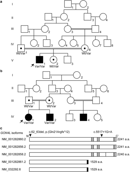
Genetic analysis of two families with homozygous GON4L variants.Pedigree of Family 1 (a) and Family 2 (b). Wt wild-type, Var variant allele. Black dots indicate a GON4L variant carrier. Arrows indicate probands. c Human GON4L isoforms with the previously reported canonical splice variant (c.5517+1G>A) which is also identified in Family 2 and the frameshift variant identified in Family 1 [c.62_63del, p.(Gln21Argfs12*)] in NM_001282860.2 are indicated. The black bar in NM_001282858.2 indicates the lack of p.Ala1958. The short isoforms (NM_001282861.2 and NM_032292.6) have the same amino acid sequence from the first methionine to p.Gln1490 as the long isoforms, and the black boxes indicate the unique regions of the short isoforms. The gray boxes indicate paired amphipathic helix 1 (PAH1, 1624–1696 amino acids), PAH2, (1706–1777 amino acids), and Myb-like (2148–2201 amino acids) domains from N- to C-terminus, respectively, based on UniProtKB (Q3T8J9). |
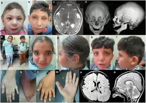
Clinical features of the patients homozygous for GON4L variants.Photographs of the face of Patient 1 at the age of 10 months (a) and 9 years and 6 months (b). He showed left microphthalmia. c Brain MRI of Patient 1 at the age of 5 months showed simplified gyral pattern. d Three-dimensional cranial CT images of Patient 1 at 10 months indicated metopic craniosynostosis. e Full picture of Patient 2 (left) and 3 (right). f, g Front and side of the face of Patient 2 at the age of 13 years old. She showed asymmetric face, a high forehead, thick eyebrows, upward slanting palpebral fissures, strabismus, broad nasal root, a broad nose with a beaked tip, deviated nasal septum, a prominent left cheek, short philtrum, thin upper lip, everted lower vermillion, broad chin, and low-set ears with folded helix. h, i Front and side of the face of Patient 3 at the age of 8 years. He shows dysmorphic features similar to Patient 2. Long fingers (j) and deviated feet with pes planus (k) of Patient 2. l, m Dorsum and palma of the hand with incomplete single transverse crease of Patient 3. Brain MRI of Patient 3 at the age of 6 [Axial T1- (n) and Sagittal T2-weighted (o)]. Deep Sylvian fissure is indicated by an arrow. |
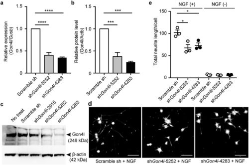
Gon4l knockdown in PC12 cells and neurite outgrowth.Knockdown efficiencies of Gon4l mRNA (a) and GON4L protein (b, c) levels in PC12 cells. mRNA levels were normalized against Gusb, and protein levels were normalized against ACTB. Data are shown as the mean ± standard error of the mean (SEM) from three independent experiments. d, e Neurite outgrowth assay in Gon4l-knockdown PC12 cells in response to NGF treatment. Representative images (d) and total neurite length in one cell (e) are shown. Dots indicate means of each independent experiment (n = 3), and the bars represent means and SEM of three independent experiments. *p ≤ 0.05, ***p ≤ 0.001, ****p ≤ 0.0001 using one-way analysis of variance (ANOVA) followed by Tukey’s multiple comparison test. |
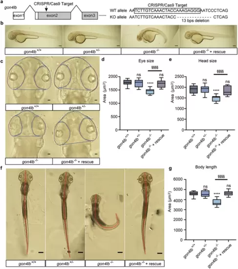
Establishment of gon4lb-null zebrafish and rescue using human GON4L mRNA.a Target design to knockout gon4lb using CRISPR/Cas9. A crRNA targeted exon 2 of gon4lb and resulted in a 13 bp deletion that generated a premature termination codon to knockout. b Representative images of 50-hpf zebrafish embryos from the gon4lb-knockout line: gon4lb+/+, gon4lb+/−, gon4lb−/−, and gon4lb−/− injected with human GON4L mRNA (rescue). All images are lateral views, with the anterior surface to the left. Scale bar: 200 µm. c Representative images of eye size and head size of 50-hpf zebrafish embryos from the gon4lb-knockout line. All images are dorsal views, with the anterior aspect at the top. The blue line delineates the contour of the head, and the pink line delineates the contour of the eye. Scale bar: 50 µm. Quantitative data showing eye (d) and head size (e) of 50-hpf zebrafish embryos: gon4lb+/+ (n = 12), gon4lb+/− (n = 12), gon4lb−/− (n = 10), and gon4lb−/− with rescue (n = 15). f Representative images of the body length of 50-hpf zebrafish embryos. All images are dorsal views, with the anterior aspect at the top. The red line indicates the position for measuring the body axis. Scale bar: 200 µm. g Quantitative data showing the body length of 50-hpf zebrafish embryos. Sample numbers are as follows: gon4lb+/+ (n = 30), gon4lb+/− (n = 30), gon4lb−/− (n = 31), and gon4lb−/− embryos with rescue (n = 17). Data are shown as the mean ± SEM; ****p ≤ 0.0001, §§§§p ≤ 0.0001 using one-way ANOVA with post hoc Tukey’s test. PHENOTYPE:
|
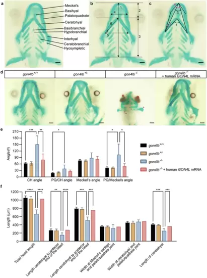
gon4lb knockout causes abnormal craniofacial development.a Craniofacial bone structure of zebrafish larvae at 5 dpf. b Measurement parameters of craniofacial bone length in wild-type zebrafish larvae: (1) Total length of the head, (2) length from the ceratohyal cartilage to the anterior end of the head, (3) length from the ceratohyal cartilage to the posterior end of the head, (4) width of the Meckel’s cartilage and palatoquadrate joint, (5) width of the ceratohyal cartilage and palatoquadrate joint, and (6) length of the ceratohyal cartilage. c Four angle parameters for craniofacial cartilage: (1) the angle between ceratohyal (CH) cartilages (CH Angle), (2) the angle between palatoquadrate (PQ) cartilage and ceratohyal cartilage (PQ/CH Angle), (3) the angle between Meckel’s cartilages (Meckel’s Angle), and (4) the angle between palatoquadrate cartilage and Meckel’s cartilage (PQ/Meckel’s Angle). All images are ventral views with the anterior at the top. Scale bars: 100 µm. d Representative images of the craniofacial cartilage of 5 dpf zebrafish larvae stained with Alcian blue and Alizarin Red (10 embryos each). The magenta-colored arrows in the panel of gon4lb−/− represent the abnormal craniofacial cartilages. All images are ventral views with the anterior at the top. Scale bar: 100 µm. e Quantitative data showing the angles of four different mineralized craniofacial cartilage elements 5 dpf zebrafish larvae. f Quantitative data showing the lengths of six different mineralized craniofacial cartilage elements in 5 dpf zebrafish larvae. Data are represented as mean ± SEM; *p ≤ 0.05; **p ≤ 0.01; ***p ≤ 0.001; ****p ≤ 0.0001 using two-way ANOVA with Tukey’s multiple comparisons test (α = 0.05). CH ceratohyal, PQ palatoquadrate. |

ZFIN is incorporating published figure images and captions as part of an ongoing project. Figures from some publications have not yet been curated, or are not available for display because of copyright restrictions. |
|
ZFIN is incorporating published figure images and captions as part of an ongoing project. Figures from some publications have not yet been curated, or are not available for display because of copyright restrictions. PHENOTYPE:
|
|
ZFIN is incorporating published figure images and captions as part of an ongoing project. Figures from some publications have not yet been curated, or are not available for display because of copyright restrictions. PHENOTYPE:
|
|
ZFIN is incorporating published figure images and captions as part of an ongoing project. Figures from some publications have not yet been curated, or are not available for display because of copyright restrictions. PHENOTYPE:
|
|
ZFIN is incorporating published figure images and captions as part of an ongoing project. Figures from some publications have not yet been curated, or are not available for display because of copyright restrictions. PHENOTYPE:
|
|
ZFIN is incorporating published figure images and captions as part of an ongoing project. Figures from some publications have not yet been curated, or are not available for display because of copyright restrictions. PHENOTYPE:
|