- Title
-
Conserved expression of the zebrafish syt4 gene in GABAergic neurons in the cerebellum of adult fishes revealed by mammalian SYT4 immunoreactive-like signals
- Authors
- Shiao, M.S., Liu, S.T., Siriwatcharapibool, G., Thongpradit, S., Khunpanich, P., Tong, S.K., Huang, C.H., Jinawath, N., Chou, M.Y.
- Source
- Full text @ Heliyon
|
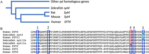
Homology of syt4 with homologs in other vertebrate species. (A) Simplified phylogenetic tree of syt4 and syt genes in zebrafish and other vertebrate species including rodents and humans. The complete phylogenetic trees were shown in Supplementary Fig. 1. (B) Alignment of partial peptide sequences of domain C2A, which has 5 calcium binding site labeling 1–5 in the figures. We included several homologs from human (SYT1-3) with no mutation at the third position of calcium biding site to compare with SYT4 and another homolog (SYT11) with substitution. The substitution of calcium biding (aspartic acid (D) to serine (S)) is conserved in zebrafish gene syt4, syt11a and syt11b (third amino acid, red box). We observed an extra substitution at the fifth binding site in zebrafish syt4 (aspartic acid (D) to glutamic acid (E), light blue box) and human SYT11. Conserved amino acids between different species were labeled in different color for an easy visualization. |
|
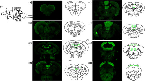
SYT4 is predominantly expressed in cerebellum in adult zebrafish. The corresponding location of each section (A–H) were indicated in (I). TeO: tectum opticum; TL: torus longitudinalis; Pit: pituitary; Val: lateral division of valvula cerebelli; Vam: medial division of valvula cerebelli; CCe: corpus cerebelli, EG: eminentia granularis; Lca: lobus caudalis of cerebellum. Green color represents mSYT4-ir signals. EXPRESSION / LABELING:
|
|
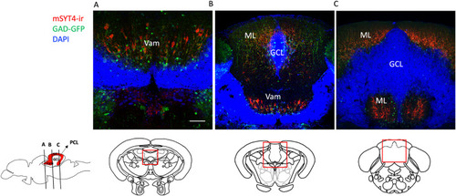
SYT4 is specific to GABAergic neurons in adult zebrafish. (A) Co-localization of mSYT4-ir signals (red) and GAD (green, a marker of GABAergic neurons) in Vam of cerebellum. (B) Co-localization of SYT4 and GAD in ML and Vam. (C) Co-localization of mSYT4-ir signals and GAD in ML. Relative locations of sections were indicated in the sagittal view of the brain illustration on the left (red color indicates ML in cerebellum). Vam: medial division of valvula cerebelli; ML: molecular layer; GCL: granule cell layer. EXPRESSION / LABELING:
|
|
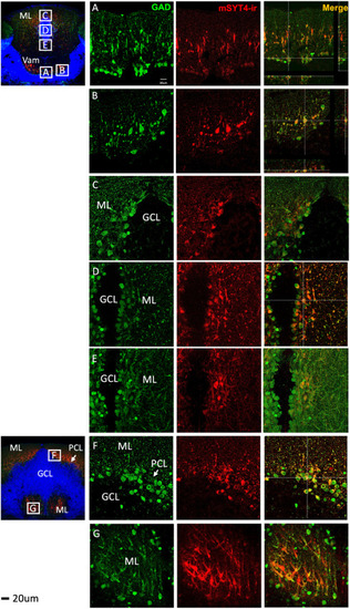
Co-localization of GAD (green) and mSYT4-ir signals (red) in the cell bodies and dendrites in different regions of the adult cerebellum. (A and B) GAD and mSYT4-ir signals in Vam. (C–E) GAD and SYT4 in CCe. Signals were only observed in the cell bodies and dendrites of the molecular layer (ML) but not in the granule cell layer (GCL). (F and G) GAD and mSYT4-ir signals in ML. The basal layer of ML is the Purkinje cell layer (PCL) indicated by the arrows. Relative locations of sections were indicated in the sagittal view of the brain illustration on the left (red color indicates ML in the cerebellum). EXPRESSION / LABELING:
|
|
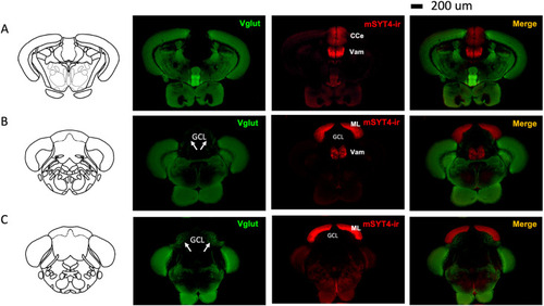
No co-localization of Vglut (glutamatergic neurons) was observed with SYT4 in the cerebellum. (A–C) Overview of ChAT and mSYT4-ir signals in the cerebellum. The strong signals of Vglut were seen in the granule cell layer (GCL) indicated by arrows. Relative locations of sections were indicated in the sagittal view of the brain illustration on the left (red color indicates ML in the cerebellum). Vam: medial division of valvula cerebelli; CCe: corpus cerebelli; ML: molecular layer; GCL: granule cell layer. EXPRESSION / LABELING:
|
|
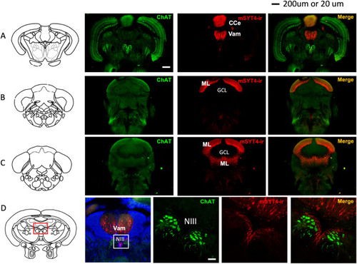
No co-localization of ChAT (cholinergic neurons) was observed with SYT4 in the cerebellum. (A–C) Overview of ChAT and mSYT4-ir signals in the cerebellum. (D) Signals of ChAT and SYT4 in oculomotor neurons (NIII). Relative locations of sections were indicated in the sagittal view of the brain illustration on the left (red color indicates ML in the cerebellum). Vam: medial division of valvula cerebelli; CCe: corpus cerebelli; ML: molecular layer; GCL: granule cell layer. EXPRESSION / LABELING:
|

