
Fig. 4
- ID
- ZDB-FIG-240611-37
- Publication
- Shiao et al., 2024 - Conserved expression of the zebrafish syt4 gene in GABAergic neurons in the cerebellum of adult fishes revealed by mammalian SYT4 immunoreactive-like signals
- Other Figures
- All Figure Page
- Back to All Figure Page
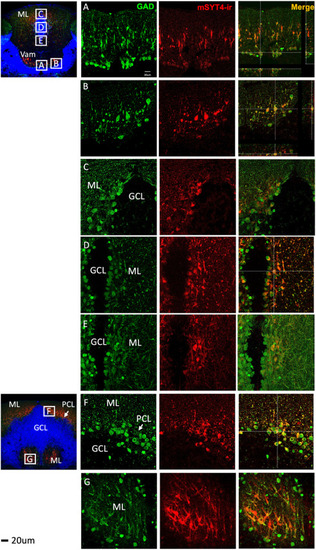
Co-localization of GAD (green) and mSYT4-ir signals (red) in the cell bodies and dendrites in different regions of the adult cerebellum. (A and B) GAD and mSYT4-ir signals in Vam. (C–E) GAD and SYT4 in CCe. Signals were only observed in the cell bodies and dendrites of the molecular layer (ML) but not in the granule cell layer (GCL). (F and G) GAD and mSYT4-ir signals in ML. The basal layer of ML is the Purkinje cell layer (PCL) indicated by the arrows. Relative locations of sections were indicated in the sagittal view of the brain illustration on the left (red color indicates ML in the cerebellum). |
| Antibody: | |
|---|---|
| Fish: | |
| Anatomical Terms: | |
| Stage: | Adult |