- Title
-
Deubiquitination of Ci/Gli by Usp7/HAUSP Regulates Hedgehog Signaling
- Authors
- Zhou, Z., Yao, X., Li, S., Xiong, Y., Dong, X., Zhao, Y., Jiang, J., Zhang, Q.
- Source
- Full text @ Dev. Cell
|
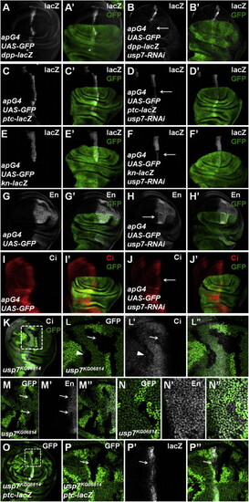
Loss of usp7 Represses Hh Signaling and Decreases Ci Protein Level All wing imaginal discs shown in this study were oriented with anterior to the left and ventral up. (A and A′) UAS-GFP (green) marks the apG4-mediated gene expression pattern. apG4 drives UAS transgenes to be specifically expressed in the dorsal region of wing discs. (B and B′) Knockdown of usp7 by apG4 attenuated the expression of dpp-lacZ (arrow). (C–J′) Knockdown of usp7 with apG4 attenuated the expression of ptc-lacZ (compare D–D′ with C–C′), kn-lacZ (compare F–F′ with E–E′), En (compare H–H′ with G–G′), and Ci (compare J–J′ with I–I′). Arrows indicate the decrease of ptc-lacZ, kn-lacZ, En, and Ci. (K–L′′) Low (K) and high (L–L′′) magnifications of wing discs carrying usp7KG06814 clones and immunostained to show the expression of GFP (green) and Ci (red). usp7KG06814 clones are recognized by the lack of GFP. (L–L′′) Enlarged views of the region marked by dashed lines in (K). (M–N′′) Wing discs carrying usp7KG06814 clones were immunostained to show the expression of GFP (green) and En (white). The clones in A compartment near A/P boundary showed decrease of En (M–M′′, arrows), whereas the clones in P compartment did not show En decrease (N–N′′). (O–P′′) usp7KG06814 clones showed a decrease of ptc-lacZ expression (arrow). (P–P′′) are enlarged views of the region marked by dashed lines in (O). See also Figure S1. |
|
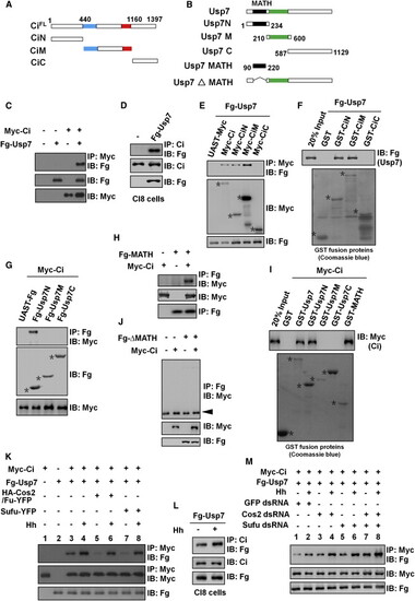
Usp7 Binds Ci through Its MATH Domain (A and B) Schematic drawings show the domains or motifs in Ci and Usp7 and their truncated fragments used in subsequent coIP and pull-down assays. Blue and red bars denote the Zn finger DNA binding and dCBP binding domains of Ci. Purple and green bars represent MATH domain and core catalytic domain of Usp7. (C) Fg-Usp7 interacted with Myc-Ci in S2 cells. (D) Fg-Usp7 interacted with endogenous Ci in Clone8 cells. (E) Fg-Usp7 associated with Myc-tagged N- and M-fragments of Ci. Asterisks indicate expressed Ci deletion mutants. (F) Extracts from S2 cells expressing Fg-Usp7 were incubated with GST or indicated GST fusion proteins. The bound proteins were analyzed by western blot. Asterisks mark GST fusion proteins. (G) Usp7 interacted with Ci through its N-region in S2 cells. Asterisks mark expressed Usp7 truncated fragments. (H) Fg-Usp7-MATH associated with Myc-Ci in S2 cells. (I) GST pull-down between Myc-tagged Ci and GST or GST-tagged Usp7 fragments. Asterisks indicate expressed GST fusion proteins. (J) MATH domain-deleted form of Usp7 did not bind Ci in S2 cells. The arrowhead indicates IgG. (K) Cos2-Fu and Sufu competed with Usp7 to bind Ci with or without Hh-conditioned medium treatment. S2 cells were treated with MG132 for 4 hr prior to harvesting the cells. (L) Hh-conditioned medium treatment promoted endogenous Ci binding to Fg-Usp7 in Clone8 cells. (M) Knockdown of cos2 or/and sufu promoted Ci binding to Usp7 in absence or in presence of Hh-conditioned medium treatment. S2 cells were treated with MG132 for 4 hr prior to harvesting the cells. See also Figures S2 and S3. |
|
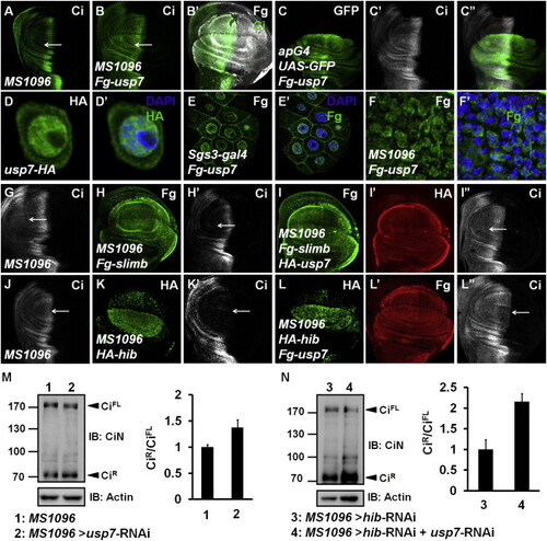
Usp7 Counteracts Ci Degradation Mediated by Both Slimb-Cul1 and Hib-Cul3 E3 Ligases UAS transgene expression with MS1096 gal4 is usually stronger in the dorsal region than in the ventral region of wing pouch. (A) A wing disc of MS1096 line was stained to show full-length Ci (green). (B and B′) A wing disc expressing Fg-usp7 with MS1096 was immunostained with Fg (white) and Ci (green) antibodies. Ci was accumulated when usp7 was overexpressed (arrow). (C–C′′) A wing disc expressing Fg-usp7 with apG4 was immunostained to show the expression of GFP (green) and Ci (white). (D and D′) Usp7-HA (green) expressed in S2 cells mainly localized in nuclei. (E and E′) Fg-Usp7 protein (green) expressed by sgs3-gal4 in salivary glands mainly localized in nuclei. (F and F′) Fg-Usp7 protein (green) expressed by MS1096 in wing discs mainly localized in nuclei. From (D) to (F′), the nuclei were marked by DAPI (blue). (G–I′′) A MS1096 wing disc (G) and the discs expressing Fg-slimb alone (H–H′) or Fg-slimb/HA-usp7 together (I–I′′) by MS1096 were immunostained to show Fg tag (green), Ci (white), and HA tag (red). Usp7 could counteract Ci degradation by Slimb-Cul1 E3 ligase (arrows). (J–L′′) A MS1096 wing disc (J) and the discs expressing HA-hib alone (K–K′) or HA-hib/Fg-usp7 (L–L′′) together by MS1096 were immunostained with HA tag (green), Ci (white), and Fg tag (red) antibodies. Usp7 could attenuate Ci degradation by Hib-Cul3 E3 ligase (arrows). (M and N) Western blot analysis of lysates from control wing discs or wing discs expressing indicated RNAi using MS1096 driver. Approximately 40 discs were dissected, lysed, and blotted with rabbit anti-CiN antibody. Signal ratio of CiR to CiFL of each lane was shown on the right. The result is presented as means ± SD of values from three independent experiments. See also Figure S4. |
|
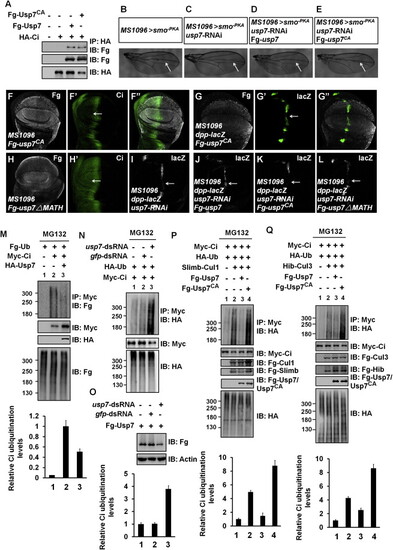
Usp7 Attenuates Slimb-Cul1- and Hib-Cul3-Mediated Ci Ubiquitination through Its Deubiquitinating Activity (A) Both Fg-Usp7 and Fg-Usp7CA could bind Myc-Ci in S2 cells. (B–E) Comparison of adult wing phenotypes from control flies (B), usp7 knockdown (C), Fg-usp7 expression in usp7 knockdown background (D), and Fg-usp7CA expression in usp7 knockdown background (E). Arrows mark the space between vein 3 and vein 4. (F–F′′) A wing disc expressing usp7CA with MS1096 was immunostained to show Fg tag (white) and Ci (green). Ci level was not upregulated by usp7CA expression (arrow). (G–G′′) A wing disc expressing usp7CA with MS1096 was stained for Fg tag (white) and dpp-lacZ (green). usp7CA overexpression decreased dpp-lacZ expression (arrow). (H and H′) A wing disc expressing usp7-ΔMATH with MS1096 was stained with Fg tag (white) and Ci (green) antibodies. Ci level was not altered by usp7-ΔMATH expression (arrow). (I–L) Wing discs expressing usp7 RNAi (I), Fg-usp7/usp7 RNAi (J), Fg-usp7CA/usp7 RNAi (K), or Fg-usp7-ΔMATH/usp7 RNAi (L) were stained for dpp-lacZ expression (white). The attenuation of dpp-lacZ caused by usp7 knockdown (I, arrow) could be restored by the expression of Fg-usp7 (J, arrow), but not Fg-usp7CA (K, arrow) and Fg-usp7-ΔMATH (L, arrow). (M) Western blots of immunoprecipitates (top) or lysates (bottom three images) from S2 cells expressing indicated proteins and treated with MG132. (N) Knockdown of usp7 using usp7-dsRNA promoted Ci ubiquitination in S2 cells. (O) usp7-dsRNA could effectively knock down Fg-usp7 expression in S2 cells. Actin acts as a loading control. (P and Q) Transfected S2 cells were treated with MG132 for 4 hr before cell harvesting. Fg-Usp7 decreased, but Fg-Usp7CA promoted Ci ubiquitination mediated by Slimb-Cul1 (P) and Hib-Cul3 (Q). From (M) to (Q), quantification analyses of the ubiquitination levels of Ci are shown below each autoradiogram. Data are means ± SD from three independent experiments. |
|
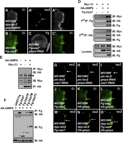
Usp7 Regulates Hh Signaling through Usp7-GMPS Complex (A–A′′) A wing disc expressing gmps RNAi by MS1096 was immunostained to show the expression of Ci (white) and dpp-lacZ (green). gmps knockdown resulted in a decrease of dpp-lacZ (arrow). (B–C′) Wing discs overexpressing usp7 alone (B) or usp7 plus gmps RNAi (C–C′) were immunostained by Fg tag (white) and Ci (green) antibodies. gmps knockdown suppressed the elevation of Ci by Usp7 expression (arrows). (D) S2 cells expressing the indicated proteins were harvested for the two-step immunoprecipitation and analyzed by western blot. (E) GMPS did not bind Ci. The indicated proteins were expressed in S2 cells. The arrowhead indicates IgG. (F) Usp7 interacted with GMPS through its M and C regions in S2 cells. Asterisks mark positions of each protein. (G–I) Wing discs expressing usp7 RNAi alone (G), gmps RNAi alone (H), or usp7 RNAi plus gmps RNAi (I) were immunostained to show ptc-lacZ expression (white). ptc-lacZ expression decreased more apparently when both usp7 and gmps were knocked down (arrows). (J–L) Wing discs overexpressing usp7 alone (J), gmps alone (K), or usp7 plus gmps (L) were immunostained for Ci expression (green). Overexpression of usp7 plus gmps resulted in a more dramatic increase of Ci (arrows). (M–O) Wing discs overexpressing usp7 alone (M), gmps alone (N), or usp7 plus gmps (O) were immunostained to show dpp-lacZ expression (green). Overexpression of usp7 plus gmps resulted in a more dramatic increase in dpp-lacZ expression (arrows). |
|
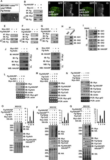
HAUSP Binds and Deubiquitinates Gli Proteins (A) Adult wing that expresses usp7 RNAi and Fg-tagged hausp. Arrow marks the space between vein 3 and vein 4. (B) Fg-HAUSP could bind Myc-Ci in S2 cells. (C and C′) A wing disc of apG4 was stained to show GFP (green) and Ci (white). (D and D′) Overexpression of hausp by apG4 resulted in an increase of Ci (white, arrow) in wing discs. (E–G) Fg-HAUSP could bind Myc-Gli1, Myc-Gli2, and Myc-Gli3 in 293T cells, respectively. The expression of corresponding proteins from cell lysate (CL) is shown below. (H) Endogenous HAUSP pulled down endogenous Gli1, Gli2, and Gli3 in 293T cells. The cells from one 10-cm plate were lysed and the lysate was equally divided into two parts for IP with control IgG or HAUSP antibody. (I) ShhN treatment increased the interaction between endogenous HAUSP and endogenous Gli proteins in 293T cells. (J and K) Sufu expression inhibited the interaction between HAUSP and Myc-tagged Gli1 (J) and Gli2 (K) in Sufu−/− MEF cells. The transfected cells were treated with MG132 for 4 hr prior to cell harvesting. (L–N) HAUSP blocked Gli protein degradation mediated by corresponding E3 ligases. 293T cells were transfected with indicated plasmids. Actin is shown as a loading control. RT-PCR analysis was performed to determine the levels of Myc-gli mRNA. The forward primer was on Myc tag, whereas the reverse primer was on gli. actin acts as a control. (O–Q) HAUSP inhibited, but HAUSPCA promoted the ubiquitination of Gli proteins mediated by E3 ligases. 293T were transfected with indicated plasmids and treated with MG132 for 4 hr before cell harvesting. Quantification analyses of the ubiquitination levels of Gli proteins are shown on right. The results were presented as means ± SD of values from three independent experiments. See also Figure S5. |
|
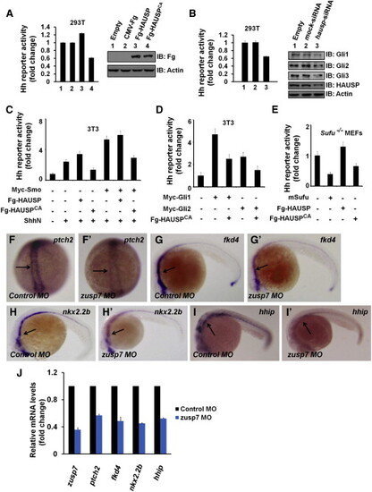
The Regulation of Usp7 on Hh Signaling Is Evolutionally Conserved in Mammalian Cells and Zebrafish (A) Gli-luciferase (Gli-luc) reporter assay in 293T cells transfected with indicated constructs. Gli luciferase activities were normalized to Renilla luciferase activities. The expression of indicated constructs was shown by western blot. (B) Gli-luc reporter assay in 293T cells transfected with indicated siRNAs. Knockdown of hausp decreased Hh pathway activity. The protein levels of HAUSP and Gli were assessed through western blotting analysis. Actin acts as a loading control. (C and D) Gli-luc reporter assay in 3T3 transfected with indicated constructs. (E) Gli-luc reporter assay in Sufu−/− MEFs transfected with indicated constructs. Of note, HAUSP regulated Hh pathway downstream of Sufu. (F–I′) Expression of Hh target genes in zebrafish embryos that were injected with the indicated MOs at 10 hpf (F–F′) or 24 hpf (G–I′). Changes in the in situ staining are marked by arrows. (J) Relative mRNA levels of zusp7, ptch2, fkd4, nkx2.2b, and hhip from zebrafish embryos indicated in (F)–(I′) are revealed by real-time PCR. See also Figure S6. |
Reprinted from Developmental Cell, 34, Zhou, Z., Yao, X., Li, S., Xiong, Y., Dong, X., Zhao, Y., Jiang, J., Zhang, Q., Deubiquitination of Ci/Gli by Usp7/HAUSP Regulates Hedgehog Signaling, 587258-72, Copyright (2015) with permission from Elsevier. Full text @ Dev. Cell

