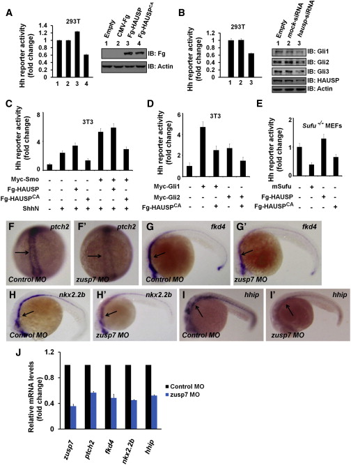
Fig. 7 The Regulation of Usp7 on Hh Signaling Is Evolutionally Conserved in Mammalian Cells and Zebrafish (A) Gli-luciferase (Gli-luc) reporter assay in 293T cells transfected with indicated constructs. Gli luciferase activities were normalized to Renilla luciferase activities. The expression of indicated constructs was shown by western blot. (B) Gli-luc reporter assay in 293T cells transfected with indicated siRNAs. Knockdown of hausp decreased Hh pathway activity. The protein levels of HAUSP and Gli were assessed through western blotting analysis. Actin acts as a loading control. (C and D) Gli-luc reporter assay in 3T3 transfected with indicated constructs. (E) Gli-luc reporter assay in Sufu−/− MEFs transfected with indicated constructs. Of note, HAUSP regulated Hh pathway downstream of Sufu. (F–I′) Expression of Hh target genes in zebrafish embryos that were injected with the indicated MOs at 10 hpf (F–F′) or 24 hpf (G–I′). Changes in the in situ staining are marked by arrows. (J) Relative mRNA levels of zusp7, ptch2, fkd4, nkx2.2b, and hhip from zebrafish embryos indicated in (F)–(I′) are revealed by real-time PCR. See also Figure S6.
Reprinted from Developmental Cell, 34, Zhou, Z., Yao, X., Li, S., Xiong, Y., Dong, X., Zhao, Y., Jiang, J., Zhang, Q., Deubiquitination of Ci/Gli by Usp7/HAUSP Regulates Hedgehog Signaling, 587258-72, Copyright (2015) with permission from Elsevier. Full text @ Dev. Cell

