
- Title
-
Impact of an irreversible β-galactosylceramidase inhibitor on the lipid profile of zebrafish embryos
- Authors
- Guerra, J., Belleri, M., Paiardi, G., Tobia, C., Capoferri, D., Corli, M., Scalvini, E., Ghirimoldi, M., Manfredi, M., Wade, R.C., Presta, M., Mignani, L.
- Source
- Full text @ Comput Struct Biotechnol J
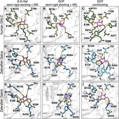
Binding modes from molecular docking and molecular dynamics simulations of the interaction of β-D-Gal and GCP with human and zebrafish GALC proteins. The active site pockets of hGALC (A,B,C), zebrafish Galca (D,E,F), and zebrafish Galcb (G,H,I) proteins are shown with the orientation of β-D-Gal (left panels) and GCP (center panels) obtained after MD simulation and for GCP obtained upon covalent docking (right panels). Proteins are shown in grey cartoon representation while residues involved in the interactions are shown in stick representation colored by element with green, cyan, or turquoise carbons for hGALC, Galca, and Galcb, respectively. β-D-Gal and GCP are shown in stick representation with carbons colored brown and magenta, respectively. H-bond interactions are shown as yellow dashed lines. The covalent bond between the ligand and the protein is shown between GCP and the neutrophile residue (E274 for human GALC, E251 and E256 for zebrafish Galca and Galcb, respectively). |

Effect of GCP on GALC activity in zebrafish. A) Extracts of 96 hpf zebrafish embryos (50 μg/sample) and of adult zebrafish brain (20 μg/sample) were incubated at room temperature with increasing concentrations of GCP in the presence of the GALC substrate LRh-6-GalcCer. After overnight incubation, the GALC product LRh-6-Cer was separated from its substrate by TLC, visualized under an ultraviolet lamp, and photographed. B) Extracts of zebrafish embryos, zebrafish, and murine adult brains, and recombinant human GALC expressed in HEK293 cells (rGALC) were incubated with GCP as in A. The GALC product LRh-6-Cer was quantified following its extraction from the silica gel plates. Data are the mean ± S.E.M of 3–4 independent experiments. C) In two independent experiments, zebrafish embryos at 1–2 cell stage were injected with 160 pmoles of GCP/embryo or vehicle. At 96 hpf, 8 GCP-treated and 8 vehicle-treaded animals were pooled and processed for GALC activity assay as in panel B. Inset: Image of the TLC analysis performed on the pools of vehicle (-) or GCP (+) treated animals. P, paired Student’s t test. D) Light sheet 3D reconstruction of Tg(neurod1:EGFP)ia50 embryos injected at 6 hpf with 160 pmoles/embryo of GCP or vehicle and visualized at 24 hpf. After image acquisition, 3D reconstructions were performed using the Arivis software (Zeiss) and exported as a single snap with the same compression settings. White arrows indicate the midbrain and eye regions of the embryos. |
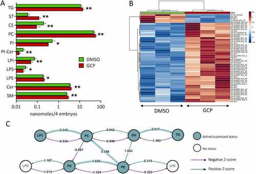
Effect of GCP on the lipid profile of zebrafish embryos. A) Levels of the different classes of lipids detected in DMSO and GCP-treated embryos. Embryos were treated at the 1–2 cell stage and lipid analysis was performed at 96 hpf. Data are expressed as nanomoles/pool of 4 embryos. * , P < 0.1; * *, P < 0.05, Student’s t test. B) Heatmap showing hierarchical clustering of the lipid species between GCP-treated zebrafish embryos and control animals (DMSO). Only the 50 most important lipids species are displayed based on their t-test p-values. Color coding represents the -Log p-value. C) Lipid subclass correlation network of GCP-treated embryos compared to controls. |
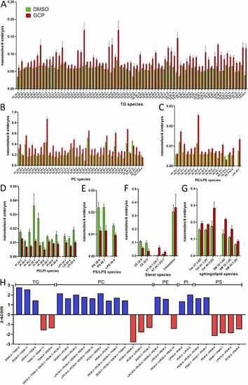
GCP affects the levels of various lipid species in zebrafish embryos. A-G) Levels of the different species of lipids whose amount was significantly affected by GCP treatment when compared to controls. Embryos were treated at the 1–2 cell stage and lipid analysis was performed at 96 hpf. Data are expressed as nanomoles/pool of 4 embryos. H) Z-score representation of the reactions that involve the most GCP-modulated species of lipids following BioPan analysis of lipidomic data. Positive z-score indicates an activated reaction, negative z-score indicates a suppressed reaction. |
|