
|
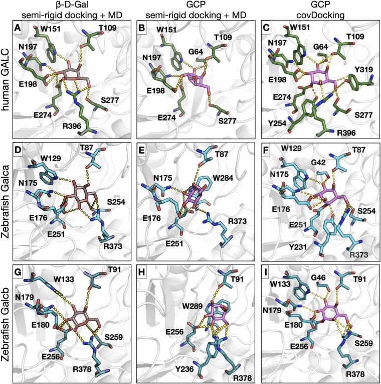
Fig. 1 Binding modes from molecular docking and molecular dynamics simulations of the interaction of β-D-Gal and GCP with human and zebrafish GALC proteins. The active site pockets of hGALC (A,B,C), zebrafish Galca (D,E,F), and zebrafish Galcb (G,H,I) proteins are shown with the orientation of β-D-Gal (left panels) and GCP (center panels) obtained after MD simulation and for GCP obtained upon covalent docking (right panels). Proteins are shown in grey cartoon representation while residues involved in the interactions are shown in stick representation colored by element with green, cyan, or turquoise carbons for hGALC, Galca, and Galcb, respectively. β-D-Gal and GCP are shown in stick representation with carbons colored brown and magenta, respectively. H-bond interactions are shown as yellow dashed lines. The covalent bond between the ligand and the protein is shown between GCP and the neutrophile residue (E274 for human GALC, E251 and E256 for zebrafish Galca and Galcb, respectively).