
- Title
-
Chronotropic and vasoactive properties of the gut bacterial short chain fatty acids in larval zebrafish
- Authors
- Sree Kumar, H., Wisner, A.S., Schiefer, I.T., Alviter Plata, A., Zubcevic, J.
- Source
- Full text @ Physiol. Genomics
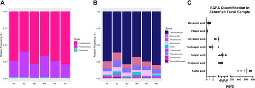
Relative abundance of bacterial taxa and composition of short-chain fatty acids (SCFAs) in adult zebrafish. A and B: 16S rRNA sequencing showing three major phyla (A) and nine genera (B) that make up the gut microbiota in the adult zebrafish (n = 6 pooled samples from 10 to 12 zebrafish/tank/sample). Bar plots show the relative abundance of bacterial taxa in each pooled sample created in RStudio. C: SCFA composition and abundance in fecal samples of adult zebrafish (n = 6 pooled samples from 10 to 12 zebrafish/tank/sample). |
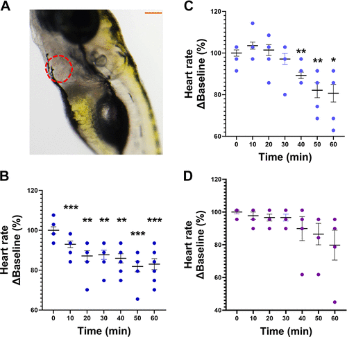
Effects of sodium butyrate, sodium acetate, and sodium propionate on heart rate (HR) in anesthetized zebrafish larvae. A: representative image showing optical clarity allowing for visualization of the heart (in red dotted lines). Scale bar in orange = 200 µm. B−D: effects of administration of 2.2 µM butyrate (B), 27.4 µM acetate (C), and 1.7 µM propionate (D) on HR in zebrafish larvae, calculated as %Baseline (0 min). Data are presented as means ± SE (n = 5–8/treatment group). One-way ANOVA with a Dunnett’s post hoc test; *P < 0.05, **P < 0.01, ***P < 0.001 |
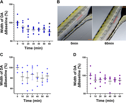
Effects of sodium butyrate, sodium acetate, and sodium propionate on vascular tone in anesthetized zebrafish larvae. A: 2.2 µM sodium butyrate produced a significant decrease in the cross-sectional width of the dorsal aorta (DA) at 60 min. B: red dotted lines mark the outline of the DA at baseline (0 min, left) and at 60 min (right) following exposure to sodium butyrate. Scale bar in orange = 200 µm. C and D: 27.4 µM acetate (C) and 1.7 µM propionate (D) produced no change. Data are presented as means ± SE and calculated as %Baseline (0 min). n = 5–8/treatment group. Repeated-measures one-way ANOVA with a Dunnett’s post hoc test; *P < 0.05. |
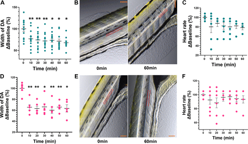
Effects of phenylephrine and angiotensin II (ANG II) on vascular tone and heart rate in anesthetized zebrafish larvae. A and D: administration of 1 µM ANG II (A) and 100 µM phenylephrine (PE) (D) produced a decrease in the cross-sectional width of dorsal aorta (DA) at all time points when compared with baseline (0 min). B and E: representative images at baseline (0 min) and the 60-min timepoint for ANG II (top) and PE (bottom), with red dotted lines marking the outline of the DA. Scale bar in orange = 200 µm. C and F: ANG II (C) and (F) PE had no effect on heart rate (beats/min). Data are presented as means ± SE (n = 6–7 PE and 6–13 ANG II) and calculated as %Baseline (0 min). Repeated-measures one-way ANOVA with a Dunnett’s post hoc test; *P < 0.05, **P < 0.01. |
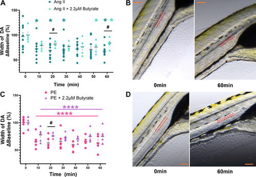
Effect of coadministration of sodium butyrate with phenylephrine (PE) and angiotensin II (ANG II) on vascular tone. A: 1 µM ANG II alone (in dark teal) significantly decreased dorsal aorta (DA) cross-sectional width at several time points compared with baseline (0 min). Coadministration of sodium butyrate (in light teal) reduced the effects of ANG II at 20 and 60 min. B: representative DA images at 0 and 60 min for ANG II, with red dotted lines marking the outline of the DA. Scale bar in orange = 200 µm. C: 100 µM PE alone (in pink) produced a significant decrease in DA cross-sectional width at all timepoints when compared with baseline (0 min). Coadministration of sodium butyrate (in purple) significantly reduced the effects of PE at 20 min. D: representative DA images at 0 and 60 min for PE, with red dotted lines marking the outline of the DA. Scale bar in orange = 200 µm. Data are presented as means ± SE and calculated as %Baseline (0 min). n = 5–10/treatment group. Two-way ANOVA with a Dunnett’s and Sidak’s multiple comparison test; *P < 0.05, ****P < 0.0001: within-group comparison to baseline (0 min). #P < 0.05 comparisons between treatment groups. |