
- Title
-
Eurycomanone stimulates bone mineralization in zebrafish larvae and promotes osteogenic differentiation of mesenchymal stem cells by upregulating AKT/GSK-3β/β-catenin signaling
- Authors
- Zhong, Y.T., Liao, H.B., Ye, Z.Q., Jiang, H.S., Li, J.X., Ke, L.M., Hua, J.Y., Wei, B., Wu, X., Cui, L.
- Source
- Full text @ J Orthop Translat
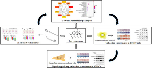
Workflow of experiments of zebrafish larvae, network pharmacology analysis, and validation experiment of mesenchymal stem cells in vitro. |

The chemical structure of EN and effects of EN on skull mineralization in zebrafish larvae. (a) Chemical structure of EN. (b) Zebrafish larvae were treated with EN (0.2, 1, and 5 μM), and skull mineralization ability was determined using ARS. (c) Mineralization areaJOTr-D-22-00514R1. (d) Mineralization IOD. (e) The mRNA expression of osteoblast-related genes (ALP, RUNX2a, SP7, and OCN) on 9-dpf in zebrafish larvae. The data are shown as the means ± SD (n = 10 zebrafish larvae/group). #P < 0.05, ##P < 0.01, ###P < 0.001 vs. Con. Scale bar: 100 μm. EN, eurycomanone; ARS, alizarin red S; IOD, integrated optical density; ALP, alkaline phosphatase, RUNX2a, runt-related transcription factor 2a; SP7, Sp7 transcription factor; OCN, osteocalcin; dpf, days after fertilization; Con, Control. (For interpretation of the references to colour in this figure legend, the reader is referred to the Web version of this article.) |
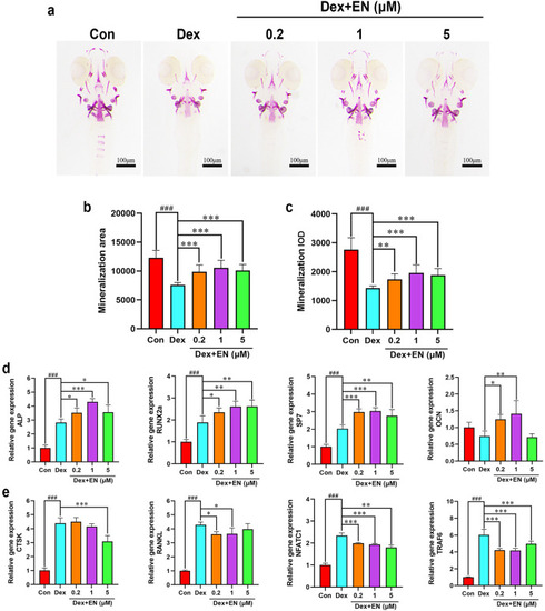
Effects of EN on Dex-induced zebrafish larvae bone loss. (a) Zebrafish larvae were treated with EN (0.2, 1, and 5 μM) combined with 10 μM Dex, and skull mineralization ability was determined using ARS. (b) Mineralization area. (c) Mineralization IOD. (d) The mRNA expression of osteoblast-related genes (ALP, RUNX2a, SP7, and OCN) on 9-dpf in Dex-induced zebrafish larvae. (e) The mRNA expression of osteoclast-related genes (CTSK, RANKL, NFATC1, and TRAF6) on 9-dpf in Dex-induced zebrafish larvae. The data are shown as the means ± SD (n = 13 zebrafish larvae/group). ###P < 0.001 vs. Con; ∗P < 0.05, ∗∗P < 0.01, ∗∗∗P < 0.001 vs. Dex. Scale bar: 100 μm. EN, eurycomanone; Dex, dexamethasone; ARS, alizarin red S; IOD, integrated optical density; ALP, alkaline phosphatase, RUNX2a, runt-related transcription factor 2a; SP7, Sp7 transcription factor; OCN, osteocalcin; CTSK, cathepsin K; RANKL, receptor of nuclear factor-κB; NFATC1, nuclear factor of activated T cells 1; TRAF, TNF receptor-associated factor 6; dpf, days after fertilization; Con, control. (For interpretation of the references to colour in this figure legend, the reader is referred to the Web version of this article.) |

Network pharmacological analysis for EN related to osteoporosis. Venn diagram of (a) EN targets and (b) OP-related genes. (c) Volcano plot of DEGs obtained from GSE35958. (d) Venn diagram of target genes of EN in OP. (e) PPI network of EN targets in OP. (f) Venn diagram of hub genes of EN in OP. (g) Target–signaling pathway/biological process interaction network of EN in OP. Note: left single node, EN; right single node, OP; middle nodes in grid arrangement, overlapping EN-OP targets; upward side nodes in circular arrangement, biological process; downward side nodes in circular arrangement, signaling pathways. (h) Biological process (BP). (i) Molecular function (MF). (j) Cellular component (CC). (k) KEGG pathway. DEGs, differentially expressed genes; EN, eurycomanone; OP, osteoporosis; PPI, protein–protein interaction; GO, Gene Ontology; KEGG, Kyoto Encyclopedia of Genes and Genomes. |
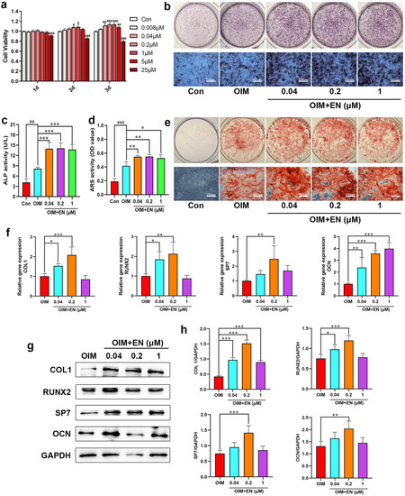
Effects of EN on cell proliferation and osteogenic differentiation in C3H10 cells. (a) Viability of C3H10 cells treated with EN at different concentrations (0.008–25 μM). (b–c) ALP staining and activity assays were measured after treating C3H10 cells with EN (0.04, 0.2, and 1 μM) for 7 days. (d–e) ARS staining and activity assays were performed after 21 days. (f) The mRNA expression levels of osteoblast-related genes (COL1, RUNX2, SP7, and OCN) in C3H10 cells were detected on day 10. (g–h) The protein expression levels of COL1, RUNX2, SP7, and OCN were detected by western blotting on day 10. #P < 0.05, ##P < 0.01, ###P < 0.001 vs. Con; ∗P < 0.05, ∗∗P < 0.01, ∗∗∗P < 0.001 vs. OIM. The data are shown as the means ± SD (n = 3). Scale bar: 100 μm. EN, eurycomanone; C3H10 cells, mouse mesenchymal stem cell line; ALP, alkaline phosphatase; ARS, alizarin red S; COL1, collagen type I; RUNX2, runt-related transcription factor 2; SP7, Sp7 transcription factor; OCN, osteocalcin; Con, Control; OIM, osteogenic induction medium. (For interpretation of the references to colour in this figure legend, the reader is referred to the Web version of this article.) |
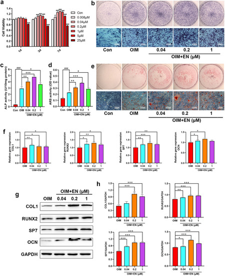
Effects of EN on cell proliferation and osteogenic differentiation in hMSCs. (a) Viability of hMSCs treated with EN at different concentrations (0.008–25 μM). (b–c) ALP staining and activity assays were measured after treating hMSCs with EN (0.04, 0.2, and 1 μM) for 7 days. (d–e) ARS staining and activity assays were performed after 28 days. (f) The mRNA expression levels of osteoblast-related genes (COL1, RUNX2, SP7, and OCN) in hMSCs were detected on day 14. (g–h) The protein expression levels of COL1, RUNX2, SP7, and OCN were detected by western blotting on day 14. #P < 0.05, ###P < 0.001 vs. Con; ∗P < 0.05, ∗∗P < 0.01, ∗∗∗P < 0.001 vs. OIM. The data are shown as the means ± SD (n = 3). Scale bar: 100 μm. EN, eurycomanone; hMSCs, human mesenchymal stem cells; ALP, alkaline phosphatase; ARS, alizarin red S; COL1, collagen type I; RUNX2, runt-related transcription factor 2; SP7, Sp7 transcription factor; OCN, osteocalcin; Con, Control; OIM, osteogenic induction medium. (For interpretation of the references to colour in this figure legend, the reader is referred to the Web version of this article.) |
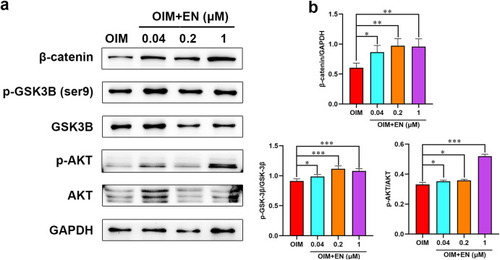
Effect of EN on AKT/GSK-3β/β-catenin pathway in C3H10 cells. (a) Protein levels of AKT/GSK-3β/β-catenin pathway in C3H10 cells were detected by western blotting on day 10: (b) β-catenin/GAPDH, p-GSK-3β (ser9)/GSK-3β, p-AKT/AKT. The data are shown as the means ± SD (n = 3). ∗P < 0.05, ∗∗P < 0.01, ∗∗∗P < 0.001 vs. OIM. EN, eurycomanone (p-) AKT (phosphorylated-) protein kinase B (p-) GSK-3β (ser9) (phosphorylated-) glycogen synthase kinase 3β at ser9; hMSCs, human mesenchymal stem cells; GAPDH, glyceraldehyde 3-phosphate dehydrogenase; OIM, osteogenic induction medium. |

Effect of EN on AKT/GSK-3β/β-catenin pathway in hMSCs. (a) Protein levels of AKT/GSK-3β/β-catenin pathway in hMSCs were detected by western blotting on day 14: (b) β-catenin/GAPDH, p-GSK-3β (ser9)/GSK-3β, p-AKT/AKT. The data are shown as the means ± SD (n = 3). ∗P < 0.05, ∗∗P < 0.01, ∗∗∗P < 0.001 vs. OIM. EN, eurycomanone (p-) AKT (phosphorylated-) protein kinase B (p-) GSK-3β (ser9) (phosphorylated-) glycogen synthase kinase 3β at ser9; hMSCs, human mesenchymal stem cells; GAPDH, glyceraldehyde 3-phosphate dehydrogenase; OIM, osteogenic induction medium. |

Effects of EN on the intervention with AKT inhibitor A-443654 in hMSCs. (a–b) ALP staining and activity assays were measured after treating hMSCs with EN (0.2 μM) and A-443654 (41 nM) for 7 days. (c–d) ARS was performed on day 28. (e) After exposed to EN and A-443654 for 14 days, the mRNA expression levels of osteoblast-related genes (COL1, RUNX2, SP7, and OCN) in hMSCs were detected. (f-g) The protein expression levels of COL1 and RUNX2 were detected by western blotting on day 14. ##P < 0.01, ###P < 0.001 vs. Con; ∗P < 0.05, ∗∗P < 0.01, ∗∗∗P < 0.001 vs. OIM; ◇P < 0.05, ◇◇P < 0.01, ◇◇◇P < 0.001 vs. A-443654; ◆P < 0.05, ◆◆P < 0.01, ◆◆◆P < 0.001 vs. EN. The data are shown as the means ± SD (n = 3). EN, eurycomanone (p-) AKT (phosphorylated-) protein kinase B; hMSCs, human mesenchymal stem cells; ALP, alkaline phosphatase; ARS, alizarin red S; COL1, collagen type I; RUNX2, runt-related transcription factor 2 (p-) GSK-3β (ser9) (phosphorylated-) glycogen synthase kinase 3β at ser9; GAPDH, glyceraldehyde 3-phosphate dehydrogenase; Con, Control; OIM, osteogenic induction medium. (For interpretation of the references to colour in this figure legend, the reader is referred to the Web version of this article.) |

Effect of EN on AKT/GSK-3β/β-catenin pathway at the present of AKT inhibitor A-443654 in hMSCs. (a) Protein levels of AKT/GSK-3β/β-catenin pathway in hMSCs were detected by western blotting on day 14: (b) β-catenin/GAPDH, p-GSK-3β (ser9)/GSK-3β, p-AKT/AKT. The data are shown as the means ± SD (n = 3). ∗P < 0.05, ∗∗P < 0.01, ∗∗∗P < 0.001 vs. OIM; ◇◇◇P < 0.001 vs. A-443654; ◆◆P < 0.01, ◆◆◆P < 0.001 vs. EN. EN, eurycomanone (p-) AKT (phosphorylated-) protein kinase B (p-) GSK-3β (ser9) (phosphorylated-) glycogen synthase kinase 3β at ser9; hMSCs, human mesenchymal stem cells; GAPDH, glyceraldehyde 3-phosphate dehydrogenase; OIM, osteogenic induction medium. |