
- Title
-
A fluidic platform for mobility evaluation of zebrafish with gene deficiency
- Authors
- Jia, X., Feng, Y., Ma, W., Zhao, W., Liu, Y., Jing, G., Tian, J., Yang, T., Zhang, C.
- Source
- Full text @ Front. Mol. Neurosci.
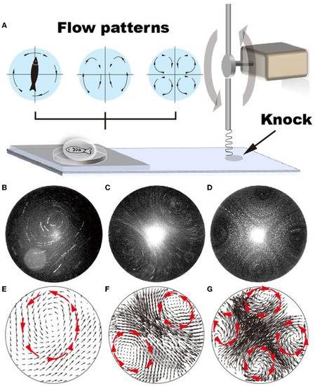
Various flow patterns can be generated by adjusting the vibration frequency of the glass substrate, i.e., from 22 to 52 Hz. (A) Schematic shows that the vibrating frequency is regulated by repeatedly knocking the glass substrate. (B–G) Using GFP fluorescent particles (1 μm in diameter) as a tracker, flow patterns generated by vibrating the glass substrate at vibrating frequencies ranging from 22 to 52 Hz are plotted. (A, D) Single recirculating flow zone generated at 22–35 Hz frequency; (B, E) two recirculating flow zones generated at 36–47 Hz frequencies; (C, F) four recirculating flow zones generated at 48–53 Hz frequencies. |
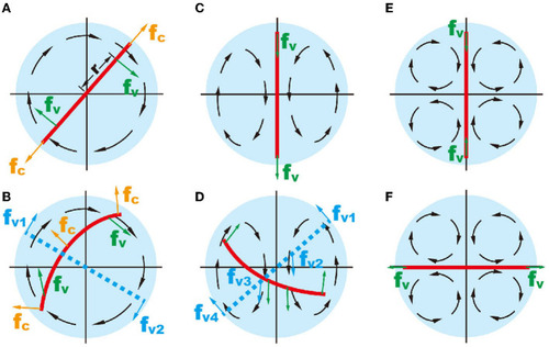
Schematic showing the mechanical forces on an inextensible object (red line) in different flow patterns. (A, B) The object in one recirculating flow zone. (C, D) two recirculating flow; and (E, F) four recirculating flow. In (B, D), the red solid lines represent a deformable object and the blue dashed lines represent an unbendable object. |
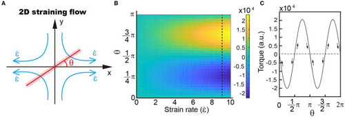
(A) Schematic showing the simplified model of four recirculating flow patterns. (B) Distribution of torques with different strain rates and object angles with respect to the outwards flow shown in a. Our results demonstrate that the torque distribution depends on the angle rather than the strain rate. (C) Variations in torque when the object rotates within the velocity field. |
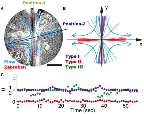
(A) Four recirculating flow zones were generated surrounding the zebrafish. It is demonstrated that there are two balanced positions, i.e., position-1 denotes the inward flow and position-2 denotes the outward flow. (B) Position distribution of different types of zebrafish shows that high-mobility fishes can be easily distinguished from the low-mobility group, i.e., type I (purple), type II (red), and type III (green) zebrafish. Each line reflects the average angle of individual zebrafish over a time span of 5 min. (C) Traces of single zebrafish's movement indicate that different types of zebrafish can be distinguished from each other. Each dot represents the position angle of a zebrafish at different time points. The scale bar denotes 1 mm. |
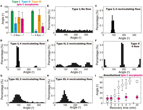
Position of zebrafish in the vibrating droplet. (A) Positions of different types of zebrafish indicate that in the droplet with four recirculating flow zones, zebrafish with disrupted mobility (i.e., type II and the ones with lipin-1 deficiency) can be better distinguished from the wide type ones (i.e., type I) as compared to the two recirculating flow conditions. (B–H) Position (i.e., angle) distribution of different types of zebrafish in various flow patterns including two and four recirculating flow patterns. (I) In the four recirculating flow patterns, anesthetized zebrafish gradually regains mobility and moves from 0° to 90° within a time span of 20 min. In contrast, zebrafish with lipin-1 deficiency remain immobile throughout the experiments. In (B), each zebrafish was maintained in the droplet for 20 min at 100% RH and 28.5°C to ensure that the random movement of zebrafish is recorded. Movements of 5–10 zebrafish are individually monitored in droplets with various flow patterns and also more than 50 zebrafish in droplets with no flow. |
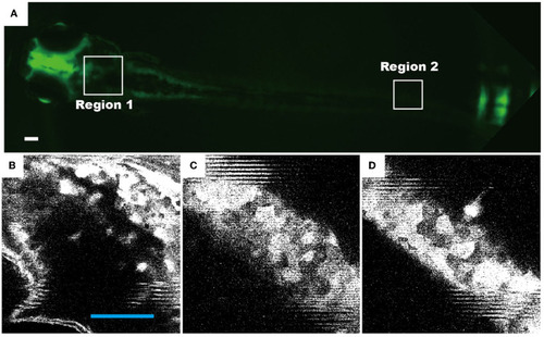
Representative fluorescence images of live zebrafish, which were transfected and maintained in a droplet of four recirculating flow patterns. The neuronal cells of transgenic zebrafish permanently express the HuC-GFP minigene (10.7 kb). Fluorescence images of 72 hpf living zebrafish: (A) as a whole fish, (B) the head region [region 1 as indicated in (A)], and (C, D) the tail region (region 2). Scale bars in all figures denote 100 μm. |