
- Title
-
Novel Compound Heterozygous Variations in MPDZ Gene Caused Isolated Bilateral Macular Coloboma in a Chinese Family
- Authors
- Zhang, S., Zhang, F., Wang, J., Yang, S., Ren, Y., Rui, X., Xia, X., Sheng, X.
- Source
- Full text @ Cells
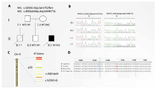
Validation of the mutation. (A) Pedigree information. In this pedigree, filled symbols indicate affected patients with MC. The unfilled symbols indicate unaffected individuals. An oblique line indicates a deceased person. Squares denote males, and circles denote females. M1 represents the c.5255C>G variant (p.Ser1752Ter), and M2 represents the c.4301delA variant (p.Asp1434fs*3). Proband (II:3) is indicated by an arrow. It shows a novel compound heterozygous MPDZ variation, c.4301delA (p.Asp1434fs*3) and c.5255C>G (p.Ser1752Ter). (B) Sanger sequencing identifying the breakpoints at c.4301delA (p.Asp1434fs*3) and c.5255C>G (p.Ser1752Ter). (C) MPDZ is localized on autosome 9p23 and contains 47 exons. (D) Arginine at positions 1752 and 1434 is highly conserved among different species according to proteomic conservation analysis. |
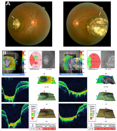
Clinical observations. (A) Fundus photograph of the proband (II:3) exhibiting prominent bilateral atrophy in the macula (white arrows); the atrophy presents well-circumscribed borders, total chorioretinal maldevelopment with a white appearance, and increased backscatter around the lesion due to the bare sclera. (B) OCT scan of the proband (II:3) showing a cup-shaped lesion with the complete absence of retina and choroid. There is a sharp margin, with significant excavation in the foveal region of both eyes. |
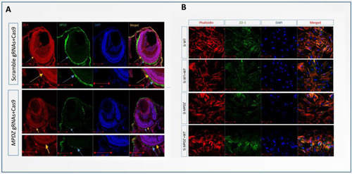
(A) IF staining showing that MPDZ localization in the ellipsoid zone of the MPDZ crispants retina, with intense green immunostaining (blue arrow), was decreased compared to the wildtype. ZO-1, mainly at the outer limiting membrane (OLM) (yellow arrow), was less expressed following the knockdown of MPDZ. (B) MPDZ knockdown with siRNA in cultured human RPE. IF staining showing the inhibition of MPDZ leading to cytoskeleton rearrangement and reduced ZO-1 expression. |

Experiment on zebrafish. (A) MPDZ crispant zebrafish displayed reduced eye area and body length compared to wildtype zebrafish. The phenotype of the MPDZ-knockdown crispants was more obvious compared to that of the wildtype. (B) Phenotypic ratio indicating the percentage of fish that uniformly showed reduced eye area and body length. The percentage of phenotypic positive fish in the MPDZ gRNA group (n = 71) was significantly greater than that in the WT gRNA group (n = 65) (p < 0.05). (C) Histological examination showing a series of phenotypes, including a small eye and a thin retina. |
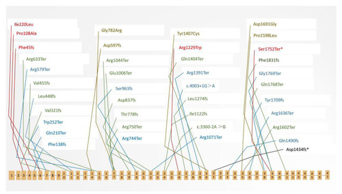
MPDZ pathogenic variations in humans. Schematic representation of the human MPDZ protein showing the distribution of variations. Blue represents a variation related to the hydrocephalus phenotype in Clinvar (https://www.ncbi.nlm.nih.gov/clinvar/, accessed on 20 August 2022). Green indicates that the pathogenic condition of the variation was “not provided” in Clinvar. Red and brown indicate that the variation was reported with the phenotype of retinitis pigmentosa (RP) and Leber congenital amaurosis (LCA), respectively. Asterisks denote variants related to macular coloboma (MC) in the present study. |