
- Title
-
CORRECTION: The Leukemia-Associated Mllt10/Af10-Dot1l Are Tcf4/β-Catenin Coactivators Essential for Intestinal Homeostasis
- Authors
- Mahmoudi, T., Boj, S.F., Hatzis, P., Li, V.S., Taouatas, N., Vries, R.G., Teunissen, H., Begthel, H., Korving, J., Mohammed, S., Heck, A.J., Clevers, H.
- Source
- Full text @ PLoS Biol.
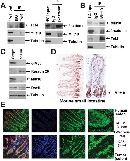
(A) Cell lysates from purified crypt fractions were immunoprecipitated with antibodies directed against endogenous Tcf4, β-catenin, and (B) Mllt10/Af10 as indicated and analyzed by Western blotting with the indicated antibodies. (C) Western blot analysis of mechanically fractionated crypt and villus from mouse small intestine using antibodies directed against a gene expressed specifically in the crypt (c-Myc), villus (Keratin 20), Tubulin as control, as well as Mllt10/Af10 and Dot1l. (D) Mllt10/Af10 antibody staining on mouse small intestinal epithelium. Arrow indicates expression in the crypt proliferative compartment. (E) Confocal image for MLLT10/AF10 (green), E-Cadherin (red), and the nuclei counter stained with DAPi (blue). MLLT10/AF10 is expressed in nuclei of normal colon epithelium in a gradient concentrated at the crypt bottom and in colorectal cancer cells. |

Cell lysates from Ls174T CRCs were immunoprecipitated with antibodies against endogenous MLLT10/AF10 (A) and DOT1L (B) complexes and analyzed by Western blotting with the indicated antibodies. (C) MLLT10/AF10 interaction with TCF4 is mediated by β-catenin. Western blot analysis of β-catenin depletion in Ls174T cells expressing doxycycline (Dox)-inducible β-catenin shRNA. Immunoprecipitated TCF4-protein complexes from untreated or Dox-treated cells were resolved by SDS-PAGE followed by Western blotting with the indicated antibodies. (D) Schematic representation of the human |

Wnt-induced association of MLLT10/AF10-DOT1L with and regulation of Wnt target genes in HEK293T cells. (A–F) ChIP assays in HEK293T cells uninduced or induced with Wnt3A conditioned media at 2 and 12 h using antibodies specific for TCF4 (A), β-catenin (B), MLLT10/AF10 (C), DOT1L (D), H3K79 di-methyl (E), and H3K79 tri-methyl (F). The immunoprecipitated DNA was analyzed by qPCR using primers specific for the |
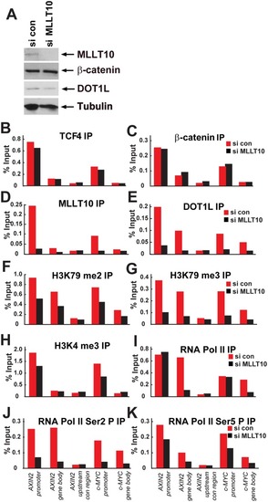
(A) Expression levels of MLLT10/AF10, β-catenin, DOT1L, and Tubulin analyzed by Western blotting after siRNA depletion of MLLT10/AF10. ChIP experiments in Ls174T CRCs containing or depleted of MLLT10/AF10 by siRNA using antibodies against (B) TCF4, (C) β-catenin, (D) MLLT10/AF10, (E) DOT1L, (F) H3K79 dimethyl, (G) H3K79 trimethyl, (H) H3K4trimethyl, (I) RNA Pol II, (J) RNA Pol II Ser2P, and (K) RNA Pol II Ser5P. Immunoprecipitated DNA was analyzed by qPCR using primers specific for |