
|
Fig 1
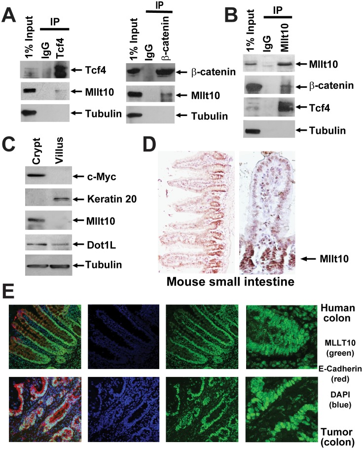
(A) Cell lysates from purified crypt fractions were immunoprecipitated with antibodies directed against endogenous Tcf4, β-catenin, and (B) Mllt10/Af10 as indicated and analyzed by Western blotting with the indicated antibodies. (C) Western blot analysis of mechanically fractionated crypt and villus from mouse small intestine using antibodies directed against a gene expressed specifically in the crypt (c-Myc), villus (Keratin 20), Tubulin as control, as well as Mllt10/Af10 and Dot1l. (D) Mllt10/Af10 antibody staining on mouse small intestinal epithelium. Arrow indicates expression in the crypt proliferative compartment. (E) Confocal image for MLLT10/AF10 (green), E-Cadherin (red), and the nuclei counter stained with DAPi (blue). MLLT10/AF10 is expressed in nuclei of normal colon epithelium in a gradient concentrated at the crypt bottom and in colorectal cancer cells.