FIGURE SUMMARY
Title

Dual-Specificity Phosphatase 14 Regulates Zebrafish Hair Cell Formation Through Activation of p38 Signaling Pathway

Authors
Wei, G., Zhang, X., Cai, C., Sheng, J., Xu, M., Wang, C., Gu, Q., Guo, C., Chen, F., Liu, D., Qian, F.
Source
Full text @ Front. Cell. Neurosci.

The phylogenetic and expression analysis of zebrafish dusp14. (A) A phylogenetic tree was generated using the PhyML software base on amino acid sequences. The pictures are mouse, giant panda, domestic cat, human, big brown bat, Chinese horseshoe bat, sperm whale, sea lion, bottlenose dolphins, lizards, zebrafish, Saccharomyces cerevisiae, and nematodes. (B,C) WISH with the dusp14 probe in wild-type zebrafish showed that the gene is expressed in the otic vesicle and lateral line at 48 hpf (B) and 72 hpf (C). (B’,C’,C”) Are the detailed information with higher magnification of the (B,C), respectively.

EXPRESSION / LABELING:
Gene:
Fish:
Anatomical Terms:
Stage Range: Long-pec to Protruding-mouth

Figure 2. Knockdown of dusp14 gene affects the hearing-related behavioral response in zebrafish. (A) Working diagram of acoustic or vibrational startle test after dusp14-MO injection at 5 dpf. (B) The distance moved of zebrafish within 1 s after the percussion stimulation. (C) Statistics of the moving rate of zebrafish during the total test. (D) The schematic diagram shows the startle response testing equipment. (E) The swimming trajectory of the control, dusp14 morphants and dusp14-mRNA and MO. (F,G) Swimming distance and peak velocity of zebrafish larvae at 5 dpf that reflected the auditory function of zebrafish larvae by examining the startle response. Values with **** above the bars are significantly different (P < 0.0001), ns means no significance.

PHENOTYPE:
Fish:
Knockdown Reagent:
Observed In:
Stage: Day 5

Knockdown dusp14 expression by morpholino decreases the number of hair cell and neuromasts in zebrafish. (A) Schematic diagram of zebrafish neuromast and inner ear hair cell. (B) Confocal imaging analysis of crista hair cells in the otic vesicle of control and dusp14 deficiency zebrafish at 72 hpf. (B′) The statistical analysis of the numbers of inner ear crista hair cells in the control and dusp14 morphants at 72 hpf. (B′′) The statistical analysis of the numbers of hair cells in the remaining neuromast L3 in the control and dusp14 morphants at 72 hpf. (C) The imaging analysis of control and dusp14 morphants at 72 hpf in fluorescent field. Scale bar = 500 μm. (C′) Quantification of the number of unilateral hair cell clusters of control and dusp14 morphants at 72 hpf. (D)in situ hybridization of the eya1 gene specifically expressed in the neuromasts showed that the number of neuromasts in dusp14-MO zebrafish was decreased. (D′) statistics results of D. Each bar represents the mean ± SEM. Values with *, ***, and **** above the bars are significantly different (P < 0.05, P < 0.001, and P < 0.0001, respectively).

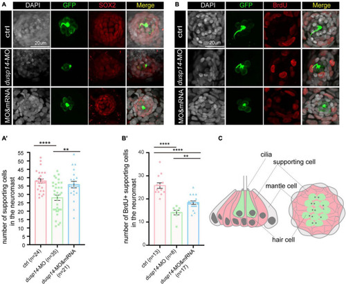
Knockdown of the dusp14 gene reduces the number of supporting cells and proliferation of supporting cells. (A) The representation of SOX2 immunofluorescence images of neuromasts in the posterior lateral line of the control and dusp14 morphants. (A′) Quantification of the number of supporting cells in the posterior lateral line neuromast of control and dusp14 mutants at 72 hpf. (B) BrdU staining for the supporting cell in the neuromasts in the posterior lateral line of the control zebrafish and dusp14 morphants. Scale bar = 20 μm. (B′) Quantification of zebrafish embryos with the BrdU+ cells in the control and dusp14 morphants. (C) Schematic diagram of the longitudinal structure and the plane structure of neuromast, the gray part is the mantle cell, the pink part is supporting cell, and the green represents hair cell. Experimental embryos were sampled at 72 hpf (n > 8). Each bar represents the mean ± SD. Values with ** and **** above the bars are significantly different (P < 0.01 and P < 0.0001, respectively).

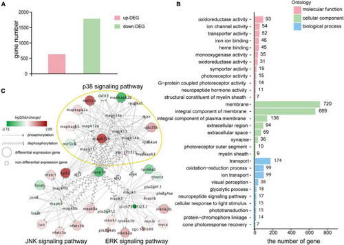
Transcriptomic sequencing data revealed p38 signaling pathways may responsible for regulation of dusp14 gene on hearing function. (A) The upregulated differentially expressed genes (DEGs) and downregulated DEGs that might be affected by knockdown dusp14 based on transcriptome data analysis. (B) Gene ontology (GO) annotation enrichment of DEGs. (C) Gene expression changes in the MAPK pathway caused by dusp14 knockdown.

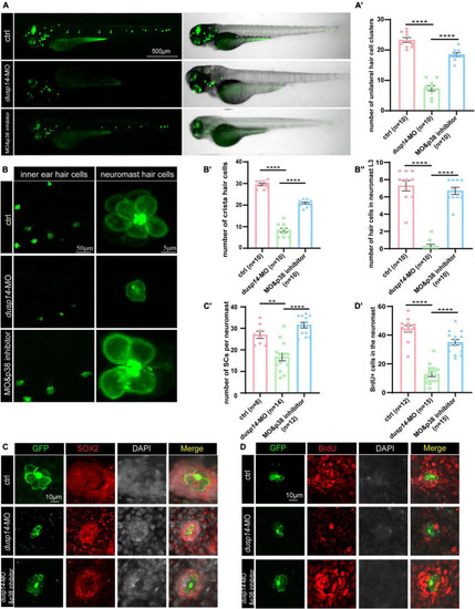
p38 signaling pathway is involved in dusp14 regulation of inner ear hearing in zebrafish. (A) The imaging analysis of control and dusp14 mutants at 72 hpf in bright field and fluorescent field. (A′) Quantification of the number of the unilateral hair cell clusters of control and dusp14 mutants at 72 hpf. (B) Confocal imaging analysis of inner ear hair cells and lateral line neuromast hair cells of control, dusp14 deficiency and dusp14-MO and p38 inhibitor coinjected zebrafish at 72 hpf. (B′) The statistical analysis of the numbers of crista hair cells for panel (B). (B′′) The statistical analysis of the numbers of hair cells in the remaining neuromast L3 for panel (B). (C) The representation of SOX2 immunofluorescence images of neuromasts in the posterior lateral line of the control, dusp14 morphants and dusp14-MO and p38 inhibitor coinjected zebrafish at 72 hpf. (C′) Quantification of the number of supporting cells per neuromast for panel (C). (D) BrdU staining for the supporting cells in the neuromasts of the control, dusp14 morphants and dusp14-MO and p38 inhibitor coinjected zebrafish at 72 hpf. (D′) Quantification of zebrafish embryos with the BrdU+ cells for panel (D). Experimental embryos were sampled at 72 hpf (n > 6). Each bar represents the mean ± SD. Values with ** and **** above the bars are significantly different (P < 0.01 and P < 0.0001, respectively).

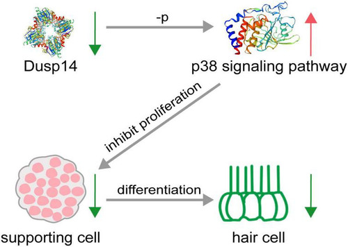
Schematic diagram of hair cell reduction caused by dusp14 knockdown. Knockdown of dusp14 expression by morpholino inhibited the proliferation of supporting cells by p38 signaling pathway, resulting in a decrease in the number of hair cells and ultimately leading to abnormal hearing and balance-related behaviors in zebrafish.

Acknowledgments
This image is the copyrighted work of the attributed author or publisher, and ZFIN has permission only to display this image to its users. Additional permissions should be obtained from the applicable author or publisher of the image. Full text @ Front. Cell. Neurosci.