FIGURE SUMMARY
Title

Sperm membrane proteins DCST1 and DCST2 are required for sperm-egg interaction in mice and fish

Authors
Noda, T., Blaha, A., Fujihara, Y., Gert, K.R., Emori, C., Deneke, V.E., Oura, S., Panser, K., Lu, Y., Berent, S., Kodani, M., Cabrera-Quio, L.E., Pauli, A., Ikawa, M.
Source
Full text @ Commun Biol

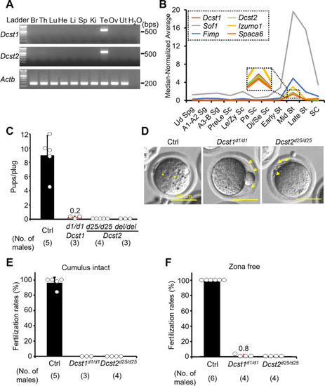
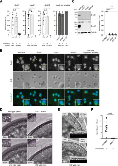
Male fertility of <italic>Dcst1</italic> and <italic>Dcst2</italic> mutant mice.

A Multi-tissue gene expression analysis. Dcst1 and Dcst2 are abundantly expressed in the mouse testis. Beta actin (Actb) was used as the loading control. Br, brain; Th, thymus; Lu, lung; He, heart; Li, liver; Sp, spleen; Ki, kidney; Te, testis; Ov, ovary; Ut, uterus. B Median-normalized level of Dcst1 and Dcst2 mRNA expression during mouse spermatogenesis. Dcst1 and Dcst2 are strongly expressed in mid-round spermatids, corresponding to other fusion-related factors. Ud Sg, undifferentiated spermatogonia; A1-A2 Sg, A1-A2 differentiating spermatogonia; A3-B Sg, A3-A4-In-B differentiating spermatogonia; Prele Sc, preleptotene spermatocytes; Le/Zy Sc, leptotene/zygotene spermatocytes; Pa Sc, pachytene spermatocytes; Di/Se Sc, diplotene/secondary spermatocytes; Early St, early round spermatids; Mid St, mid round spermatids; Late St, late round spermatids; SC, Sertoli cells. C Male fecundity. Each male was caged with 2 wild-type females for >1 month. Dcst2d25/wt and del/wt males were used as the control (Ctrl). Dcst1d1/d1, Dcst2d25/d25, and Dcst2del/del males succeeded in mating [number of plugs: 19 (Ctrl), 17 (Dcst1d1/d1), 42 (Dcst2d25/d25), 24 (Dcst2del/del)], but the females very rarely delivered pups [pups/plug: 9.0 ± 2.8 (Ctrl), 0.2 ± 0.2 (Dcst1d1/d1), 0 (Dcst2d25/d25), 0 (Dcst2del/del)]. D Egg observation after IVF. After 8 h of incubation, pronuclei were observed in the control heterozygous sperm (asterisks). However, Dcst1 KO and Dcst2 KO sperm accumulated in the perivitelline space (arrows). The scale bars are 50 μm. E Sperm fertilizing ability using cumulus-intact eggs in vitro. Dcst1 KO and Dcst2 KO sperm could not fertilize eggs [fertilization rates: 96.5 ± 7.1% (Ctrl, 231 eggs), 0% (Dcst1d1/d1, 97 eggs), 0% (Dcst2d25/d25, 197 eggs)]. F Sperm fertilizing ability using ZP-free eggs in vitro. Dcst1 KO and Dcst2 KO sperm rarely fertilized eggs [fertilization rates: 100% (Ctrl, 142 eggs), 0.8 ± 1.6% (Dcst1d1/d1, 94 eggs), 0% (Dcst2d25/d25, 88 eggs)]. All values in this figure are shown as the mean ± SD.

Adhesion and fusion ability of <italic>Dcst1</italic> and <italic>Dcst2</italic> mutant sperm to oocyte plasma membrane.

A Binding ability. Dcst1 KO and Dcst2 KO sperm could bind to the oolemma after 30 min of incubation. The scale bars are 50 μm. B Detection of IZUMO1. The band signals of IZUMO1 in TGC and sperm of Dcst1d1/d1 and Dcst2d25/d25 male mice were comparable to the control wild-type sperm. SLC2A3, one of proteins in sperm tail, was used as the loading control. C, D Acrosome status of binding sperm. Live sperm bound to the oolemma were stained with the IZUMO1 antibody, and IZUMO1 only in the acrosome reacted (AR) sperm was detected. The AR-sperm of both Dcst mutants could bind to the oolemma, and their numbers were higher than the control heterozygous sperm [3.27 ± 2.31 (Ctrl, 93 eggs), 7.34 ± 5.09 (Dcst1d1/d1, 68 eggs), 4.74 ± 2.93 (Dcst2d25/d25, 89 eggs)]. **p < 0.01. The scale bars are 25 μm. E, F Fusion ability. The ZP-free eggs pre-stained Hoechst 33342 were used for sperm-egg fusion assay. Hoechst 33342 signal transferred to control sperm heads, indicating that sperm fused with eggs (panel E, arrow). However, Dcst1 KO and Dcst2 KO sperm rarely fused with eggs [fused sperm/egg: 1.52 ± 0.35 (Ctrl, 113 eggs), 0.04 ± 0.05 (Dcst1d1/d1, 73 eggs), 0 (Dcst2d25/d25, 73 eggs)]. The scale bars are 25 μm. All values are shown as the mean ± SD.

Detection of DCST1/2 in TGC and sperm and interaction of DCST1/2.

A Rescue of male fertility. Dcst1d1/d1 males with Dcst1-3xHA Tg insertion and Dcst2d25/d25 males with Dcst2-3xHA Tg insertion were generated (Fig. S10), and their fertility was rescued [number of plugs: 17 (Dcst1d1/d1), 25 (Dcst1d1/d1;Tg), 42 (Dcst2d25/d25), 15 (Dcst2d25/d25;Tg)]. The fecundity data in Dcst1d1/d1 and Dcst2d25/d25 males is replicated from Fig. 1C. All values are shown as the mean ± SD. B Detection of DCST1 and DCST2 in TGC and sperm. The protein extract of TGC (100 μg) and sperm (6.6 × 10^6 sperm) was used for SDS-PAGE. The HA-tagged DCST1 and HA-tagged DCST2 were detected in TGC and sperm. Total proteins in the membrane were visualized by CBB staining. Triangle marks show the expected molecular size of DCST1 (about 80 kDa) and DCST2 (about 77 kDa). C Localization of DCST2 in sperm. The HA-tagged DCST2 was localized in the anterior acrosome before the acrosome reaction, and then translocated to the equatorial segment in acrosome-reacted sperm (arrows). Wild-type sperm were used as the negative control. PNA was used as a marker for the acrosome reaction. The fluorescence in the sperm tail was non-specific. The scale bars are 25 μm. D Co-IP and western blotting of the interaction between IZUMO1 and DCST1/2. The TGC and sperm lysates from Ctrl (Dcst2wt/wt and d25/wt mice), Dcst1;Tg, and Dcst2;Tg males were incubated with anti-HA tag antibody-conjugated magnetic beads, and then the eluted protein complex was subjected to western blotting. The HA-tagged DCST1 was detected only in the IP product from TGC, and the HA-tagged DCST2 was detected in the IP-product from TGC and sperm. IZUMO1 was not detected in the co-IP products. Red and blue triangle marks show the expected molecular size of DCST1 (about 80 kDa) and DCST2 (about 77 kDa), respectively. E Interaction between DCST1 and DCST2 in HEK293T cells. The protein lysate collected from HEK cells overexpressing Dcst1-3xFLAG and Dcst2-3xHA was incubated with anti-HA tag antibody-conjugated magnetic beads. The FLAG-tagged DCST1 was detected in the eluted protein complex. ADAM1B, a sperm protein that localizes to the sperm surface and is not involved in sperm-egg fusion, was used for negative control.

Binding assay between ZP-free eggs and HEK293T cells overexpressing <italic>Dcst1/2</italic>.

A Detection of DCST1/2 and IZUMO1. FLAG-tagged DCST1, HA-tagged DCST2, and 1D4-tagged IZUMO1 were detected in HEK293T cells overexpressing Dcst1-3xFLAG, Dcst2-3xHA, and Izumo1-1D4. B, C Observation of ZP-free eggs incubated with HEK293T cells overexpressing Dcst1/2 and Izumo1. The HEK293T cells overexpressing Dcst1 or Dcst2 did not attach to the oocyte membrane. Even when the HEK cells overexpressing Dcst1/2 were used for the assay, these cells failed to bind to ZP-free eggs. The HEK293T cells overexpressing Dcst1/2 and Izumo1 could bind to the oocyte membrane but could not fuse with an egg. n.s.: not significant (p = 0.28). The scale bars are 100 μm. All values are shown as the mean ± SD.

<italic>Dcst1</italic> and <italic>dcst2</italic> are essential for male fertility in zebrafish.

ADcst1 and dcst2 mutant zebrafish are male sterile. Quantification of fertilization rates as assessed by the number of embryos that progress beyond the one-cell stage. Left three panels: Males of different genotypes [wild-type sibling (+/+; white); heterozygote sibling (+/−; light gray); homozygote sibling (−/−; dark gray)] were crossed to wild-type females; right panel: homozygous mutant females (−/−; dark gray) of the indicated genotypes were crossed to wild-type males. The number of individual clutches and the total number of eggs per genotype are indicated. Data are means ± SD; adj. ****p < 0.0001 (Kruskal–Wallis test with Dunn’s multiple-comparisons test); n.s., not significant. B Dcst2 localizes to the periphery of the sperm head. Immunofluorescent and differential interference contrast (DIC) images of wild-type and mutant zebrafish sperm. Sperm were stained with DAPI (blue) to visualize nuclei and an antibody against the RING-finger domain of zebrafish Dcst2. Dcst2 localizes to distinct foci around the sperm head (green arrowheads). Dcst2 foci are reduced (dcst1−/−) or not detectable (dcst2−/−; dcst1/2−/−) in mutant sperm, while overall sperm morphology in DIC images appears normal for all genotypes. Autofluorescence of the sperm mid-piece appears as a uniform signal in the Dcst2 channel (orange arrowhead) in all genotypes. Scale bar: 5 μm. Boxed inset shows an individual enlarged sperm head for each genotype. C Dcst1 and Dcst2 are absent in mutant sperm. Exemplary immunoblot of sperm samples of wild-type and mutant genotypes probed with antibodies against zebrafish Dcst1 and Dcst2. Tubulin protein levels of the same blot are shown as loading control. For Dcst2, an unspecific band (asterisk) is detected in all genotypes at ~250 kDa. Quantification of Dcst2 levels of wild-type and mutant sperm based on n = 4 biologically independent immunoblots. Values were normalized to tubulin and then to wild-type levels. Data are means ± SD; ****p < 0.0001 (one-way ANOVA and multiple comparisons analysis). DDcst2 mutant sperm are motile and reach the micropyle. Images from time-lapse movies of wild-type (left) or dcst2−/− (right) sperm added to wild-type eggs. Sperm (magenta) were labeled with MitoTracker and added to inactivated eggs. Sperm and eggs were activated by addition of water just before the start of the movie. The micropyle (white arrow), a preformed funnel in the egg coat through which the sperm reach the oolemma, is outlined with a dashed white line. Top images depict the first acquired image following sperm addition: no sperm has entered the micropylar area in either wild-type or mutant samples. Bottom images (125 and 75 s after sperm addition in wild-type and dcst2−/− samples, respectively): sperm can readily be detected within the micropylar area (inset). Scale bar: 75 μm. E, FDcst2 mutant sperm are defective in stable binding to wild-type eggs. E Images from a time-lapse movie of wild-type (top) or dcst2−/− (bottom) sperm added to activated and dechorionated wild-type eggs. Sperm (magenta) were labeled with MitoTracker and activated at the time of addition to the eggs. Wild-type sperm show clear binding to the surface of the egg (inset), while dcst2−/− sperm are unable to stably bind to the oolemma. Scale bar: 50 μm. F Binding of sperm was assessed by quantifying the number of stably bound sperm in a 1-min time window. The number of independent experiments is indicated. ****p < 0.0001 (Mann–Whitney test).

Acknowledgments
This image is the copyrighted work of the attributed author or publisher, and ZFIN has permission only to display this image to its users. Additional permissions should be obtained from the applicable author or publisher of the image. Full text @ Commun Biol