FIGURE SUMMARY
Title

Meiotic Chromosome Dynamics in Zebrafish

Authors
Imai, Y., Olaya, I., Sakai, N., Burgess, S.M.
Source
Full text @ Front Cell Dev Biol

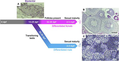
Gonadal sex differentiation and oocyte staging in zebrafish. Adapted from Blokhina et al. (2021). At 13-days post fertilization (dpf) the gonad is considered bipotential (i.e., can develop as either a testis or an ovary) and is made up of pre-follicle and early follicle stages (referred to as stage IA and IB, respectively, based on staging in Selman et al., 1993) and includes cells in meiotic prophase I. Following Stage IA oocyte formation, testis transitioning occurs ∼20–30 dpf in roughly 50% of larvae. In this case, oocytes undergo apoptosis and spermatogenesis is initiated. In animals that differentiate as females, oocytes develop further, and Stage IA oocyte production continues through adulthood. (A) Hematoxylin and Eosin (H&E) stained section of 24 dpf gonad. Scale bar = 20 μm. (B) H&E section of adult ovary. Scale bar = 100 μm. (C) H&E section of adult testis. Scale bar = 20 μm.

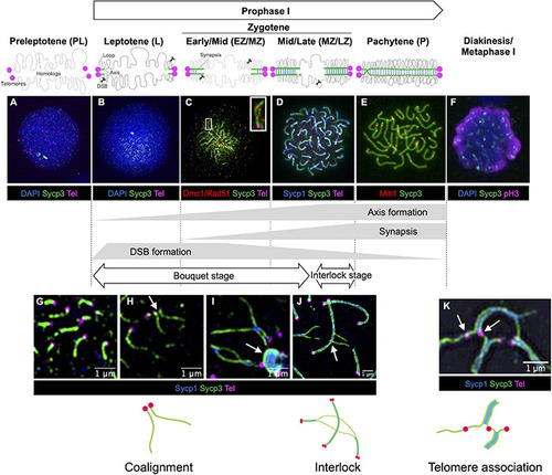
Stages of meiotic prophase I in zebrafish. Immunofluorescence staining of synaptonemal complex protein 3 (Sycp3) with telomeres (Tel), DNA (DAPI) and/or stage specific markers on zebrafish spermatocyte spreads observed by conventional immunofluorescence microscopy (A–F) and by super resolution microscopy (G–K). Diagrams of homologous chromosome pairs (gray lines) indicate axis formation (green lines) and synapsis (blue lines) from telomeres (circles in magenta). (A) In the preleptotene stage, telomeres are yet to cluster and aggregates of Sycp3 are observed. (B) In the leptotene stage, telomeres cluster in the bouquet and axis formation as seen by the formation of Sycp3 lines immediately adjacent to telomeres. (C) DSBs near telomeres in leptotene and early/mid-zygotene (EZ/MZ) stages, visualized by staining DNA recombinases (Dmc1/Rad51; adapted from Takemoto et al., 2020). A region marked as a white rectangle is shown at a higher magnification at the top right. (D) Synapsis between homologs visualized by synaptonemal complex protein 1 (Sycp1) staining in a mid- to late zygotene (MZ/LZ) nucleus. (E) In the pachytene stage, axis formation and synapsis are completed and chromosomes are aligned from end-to-end. Future crossover sites are visualized by staining of MutL homolog 1 (Mlh1), which is involved in DSB repair. (F) A phosphohistone H3 (pH3) positive nucleus with broken Sycp3 signals. (G) Axes originate from telomere regions. (H) Coaignment between homologs, as indicated by parallel segments of axes (arrow). (I) Synapsis initiates between end regions. Telomeres are often seen associated with polycomplexes made up of Sycp3 and Sycp1 proteins (arrow). (J) End-to-end synapsis can result in interlocks where one or two chromosomes (in this case two synapsed homologs) can be trapped between another synapsed pair (arrow). Interlocks are often seen with local regions of asynapsis. (K) Telomere associations can persist into zygotene (shown here) and pachytene (not shown) although their numbers are reduced. Sometimes a stretch of axis can be seen spanning the ends of two unrelated chromosomes (arrow). Schematic diagrams of chromosome configurations are shown at the bottom. In (A,B,F) blue indicates DAPI stained DNA while in (D,G–J) blue indicates Sycp1 protein. (C) Is adapted from Takemoto et al. (2020). (F) Is modified from Ozaki et al. (2011). (G–I) Were previously published in Blokhina et al. (2019). (A,B,D,E,J) Replicate previously published images.

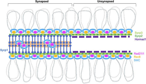
Meiotic chromosome axis structures. The synaptonemal complex comprises the lateral elements (LE, Sycp2 and Sycp3) that run along the lengths of homologous chromosomes joined by a central region that contains the transverse filament (Sycp1) and a central element (a region indicated in orange). Chromosomes joined by this tripartite structure are considered “synapsed.” Prior to synapsis, the LE is referred to as the axial elements (AE) where chromatin is organized into loops that are serially attached to the axis. The chromosome axis is made up of cohesins, the axial element proteins and HORMA-domain proteins. In zebrafish, axis localization has been observed for the cohesin components Smc3, Smc1β, and Rad21l1, the axial element proteins Sycp2 and Sycp3, and Hormad1. Localization of Rec8a/b (there are two paralogs in zebrafish) and Hormad2 (not shown) remains to be determined. Homologous chromosome pair at the ends as seen by the coalignment of axial elements. While the Rec8 cohesin complex most likely links sister chromatids together, it is not clear what DNA sequences are associated with Rad21l1 complex, however, it could play a role similar to the COH-3/4 cohesin complexes that enable the formation of asymmetric chromosome loops in C. elegans (Wolger et al., 2020).

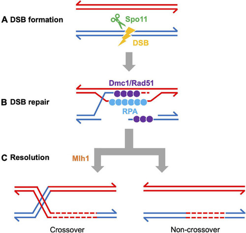
Meiotic recombination pathway showing steps mediated by proteins described in the text. Adapted from Baudat et al., 2013. Red and blue lines represent double strand DNAs of maternal and paternal origins. Broken lines indicate newly synthesized DNA strands during DSB repair. (A) DSB formation requires the topoisomerase-like protein Spo11. (B) The repair of meiotic DSBs involves strand exchange mediated by the ssDNA binding complex RPA, the recombinases Dmc1 and Rad51, and Brca2. (C) Recombination intermediates are resolved to form either crossover or non-crossover products. Crossover resolution involves Mlh1. Other proteins have also been shown to act in meiotic recombination in mammals (reviewed in Baudat et al., 2013) and in budding yeast (reviewed in Yadav and Claeys Bouuaert, 2021). Presented proteins have been studied in zebrafish by mutation and/or cytological analyses. It has not been definitively shown that FANCL/Fancl acts at these stages in both mouse and zebrafish.

Acknowledgments
This image is the copyrighted work of the attributed author or publisher, and ZFIN has permission only to display this image to its users. Additional permissions should be obtained from the applicable author or publisher of the image. Full text @ Front Cell Dev Biol