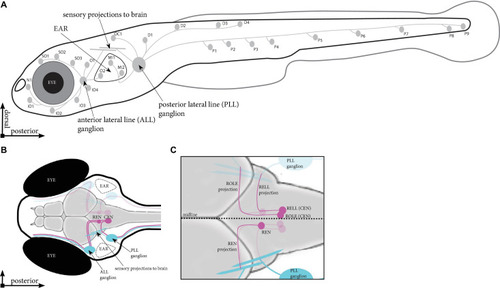
Schematic overview of the lateral line in zebrafish. (A) Location of the neuromasts in a zebrafish larva at 5 days post fertilisation (dpf). Indicated are the neuromasts identified in Tg(HGn39D), the anterior and posterior sensory ganglia and the projections they send to the neuromasts and into the brain. (B) Dorsal view of the head of a 5 dpf zebrafish larva. Indicated are the sensory ganglia and their projections (cyan) as well as the REN and CEN in the rhombencephalon (magenta), where we find the inhibitory efferent cells projecting to the lateral line. (C) Overview of the different cells in the REN and CEN (magenta). The REN contains a single cell type, which sends a projection anteriorly that turns toward the ALL ganglion. The CEN contains two cell types; ROLE, which projects past and follow the projection path of the REN cell; and a RELL cell, which projects till the REN cell, where it turns toward the PLL ganglion. ALL, anterior lateral line; PLL, posterior lateral line; REN, the rostral efferent nucleus; CEN, the caudal efferent nucleus nucleus; ROLE, the rhombencephalic octavolateral efferent neuron; RELL, the rhombencephalic efferent neuron to the lateral line.

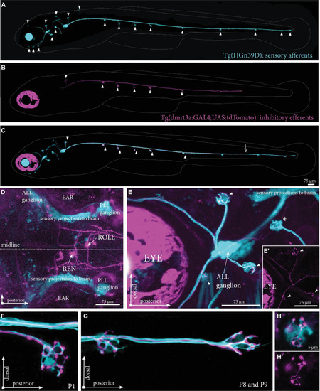
Inhibitory efferent projections to the lateral line in zebrafish larvae. (A) Overview of the inhibitory efferent projections in a Tg(dmrt3a:GAL4;UAS:RFP) 5 dpf larvae. Arrowheads indicate sites of neuromast innervation. Note that this is not the original confocal image, but a modified version to show the inhibitory efferent projections only; for the original image, please see Supplementary Figure 1. (B) Top view showing the head of the same larvae. Features marked are the eye, the ear and the olfactory bulb (OB; RFP-positive). Boxed area is shown in (C). (C) Confocal image showing the REN and CEN in the hindbrain. Arrows indicate projections to the ALL ganglion and the PLL ganglion. (D) Schematic overview to indicate sites of ALL (S01-SO3; above the eye) and PLL (P1, above the yolk) back-fills. (E) Side view of a PLL back-fill. Back-fills were performed posterior of the PLL ganglion. Note the dendritic process from the ganglion to the brain (above the ear). (F) Overlay of back-filled inhibitory efferent cells in the CEN with the cells labeled by Tg(dmrt3a:GAL4;UAS:GCaMP5g). (F’) Back-fill of ROLE and RELL cells located in the CEN. (F”) Location of overlap by back-filled cells are indicated by white dotted circles in a Nacre Tg(dmrt3a:GAL4;UAS:GCaMP5g) larva. OB, olfactory bulb; ALL, anterior lateral line; PLL, posterior lateral line; REN, the rostral efferent nucleus; CEN, the caudal efferent nucleus nucleus; ROLE, the rhombencephalic octavolateral efferent neuron; RELL, the rhombencephalic efferent neuron to the lateral line.

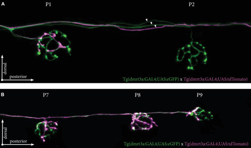
Projections by single inhibitory efferent cells. (A) Overview of the sensory afferent projections in Tg(HGn39D) at 5 dpf. Arrowheads indicate sites of neuromast innervation. (B) A single labeled inhibitory efferent neuron in Tg(dmrt3a:GAL4;UAS:tdTomato) showing partial coverage of the lateral line by its projections. Arrowheads indicate sites of neuromast innervation. (C) Overlay of A and B. Arrowheads indicate sites of afferent and efferent innervation of neuromasts. Arrow indicates the growth cone of the inhibitory efferent projection. Note that A-C do not show the original confocal image, but a modified version to show the lateral line projections only; for the original images, please see Supplementary Figure 2. (D) Top view of a Tg(dmrt3a:GAL4;UAS:tdTomato), where only a single cell in the REN and a single cell in the CEN (ROLE) can be seen. (E) Zoomed image of the ALL ganglion of Tg(HGn39D; dmrt3a:GAL4;UAS:tdTomato) reveal a single projection along the lateral line nerve of the ALL. Arrowheads indicate sites of neuromast innervation by the inhibitory efferent. Asterisks indicate neuromasts without inhibitory efferent innervation [(E’) shows the inhibitory efferent projection only]. (F–G) Higher magnification of Tg(HGn39D; dmrt3a:GAL4;UAS:tdTomato), showing the sensory afferents and a single efferent projection innervating a neuromast at P1 (F) and at P8 and P9 (G). (H) Example of a neuromast that is only partially innervated by the single inhibitory efferent [(H’) shows the inhibitory efferent projection only]. ALL, anterior lateral line; PLL, posterior lateral line; REN, the rostral efferent nucleus; ROLE, the rhombencephalic octavolateral efferent neuron; RELL, the rhombencephalic efferent neuron to the lateral line.

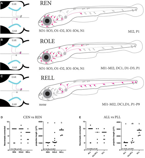
Summary of the projection paths of individual inhibitory efferent cells. (A–C) Schematic zebrafish larvae showing the cumulative projection paths of REN (n = 8), ROLE (n = 10), and RELL (n = 9) cells. Left drawing illustrates the projection paths of the REN, ROLE, and RELL cells (highlighted in magenta) within the rhombencephalon; projections of the sensory ganglion into the brain are shown in cyan. For each inhibitory efferent the cumulative projection path across the lateral line is highlighted (magenta) in the schematic zebrafish larvae on the right (for individual processes, please see Supplementary Figure 3). Identity of innervated neuromasts is listed below each schematic (left: anterior lateral line neuromasts; right: posterior lateral line neuromasts). (D) Quantification of the number of neuromasts connected revealed no significant differences between REN, ROLE, and RELL cells (K = 2.789, p = 0.25). The total length of the projections made by the RELL cells was significantly longer compared to that of the REN and ROLE cells (K = 18.59, p < 0.0001). (E) No significant differences were found between the number of neuromasts connected by individual inhibitory efferent cells that project to the ALL only, ALL and PLL, or PLL only (K = 5.609, p = 0.06). The total length of the projections made by cells that connect in the PLL was significantly longer compared to those connecting to the ALL but not the ALL+PLL (K = 16.84, p < 0.0001). Age of larvae: 5dpf. ALL, anterior lateral line; PLL, posterior lateral line; REN, the rostral efferent nucleus; CEN, the caudal efferent nucleus nucleus; ROLE, the rhombencephalic octavolateral efferent neuron; RELL, the rhombencephalic efferent neuron to the lateral line.

Time-lapse recordings of sensory afferent and inhibitory efferent processes growing along the PLL. (A–D) Time frames of time-lapse imaging made by digital light-sheet microscopy in a Tg(HGn39D; dmrt3a:GAL4;UAS:tdTomato) larva. Arrowheads indicate the growth cone for the sensory afferent projection and arrows indicate the growth cone of the inhibitory efferent projection. Asterisks indicates projections on the other side of the larvae’s body visible in the maximum intensity projections. (E) The growth rate between the sensory afferent projections (1.15 ± 0.18 μm/min) and inhibitory efferent projections (1.0 ± 0.17 μm/min) was not significantly different (t = 1.581, df = 14, p = 0.14). (F–I) Higher magnification time-lapse confocal imaging of the synaptic terminals of afferent processes and a single inhibitory efferent process [(F’–I’) show inhibitory efferent projection only]. Recordings show innervation of a neuromasts by sensory afferents prior to inhibitory efferent innervation. Arrows indicate the growth cone of the inhibitory efferent. Age of larva in (A) 36 hpf; Age of larva in (F) is 54 hpf.

Tg(dmrt3a:GAL4;UAS:tdTomato) injected with Kaloop plasmid reveal multiple inhibitory efferent cells innervating a single neuromast. (A) One-cell stage embryos of Tg(dmrt3a:GAL4;UAS:tdTomato) were injected with Kaloop plasmid to generate larvae with sparsely labeled tdTomato and eGFP inhibitory efferents. (B) Schematic overview of the area shown in (E). (C–D) Confocal images showing the view of sparsely labeled inhibitory efferent cells in their nuclei. A ROLE (tdTomato-positive; magenta) and a RELL (eGFP-positive; white) cell were identified (C) and their projections traced across the lateral line (D) dorsal view and (E) lateral view of the inhibitory efferent projections at the level of the ear. In this case, both efferent cells were connected to the same neuromast located on the ear (MI1) [(E’) depicts the MI1 neuromast at higher magnification]. Age of larvae: 5 dpf. ROLE, the rhombencephalic octavolateral efferent neuron; RELL, the rhombencephalic efferent neuron to the lateral line. (A) was partially created with BioRender.com

Tg(dmrt3a:GAL4;UAS:tdTomato) crossed with Tg(dmrt3a:GAL4;UAS:eGFP) reveal multiple inhibitory efferent processes innervating a single neuromast. (A) Confocal images of neuromast P1 and P2 of the PLL. All efferent projections were labeled by Tg(dmrt3a:GAL4;UAS:eGFP) (green) and single efferent projection was labeled by Tg(dmrt3a:GAL4;UAS:tdTomato) (magenta). Note how the single tdTomato-positive (magenta) projection connects to the P1 neuromast, but skips the P2 neuromast. Arrowheads indicate three different efferent projections along the PLL. (B) In the same larvae as in (A), the single tdTomato-positive (magenta) projection connects to the last three neuromasts of the PLL (P7-P9). Age of larvae: 5 dpf.

Acknowledgments
This image is the copyrighted work of the attributed author or publisher, and ZFIN has permission only to display this image to its users. Additional permissions should be obtained from the applicable author or publisher of the image. Full text @ Front. Neuroanat.