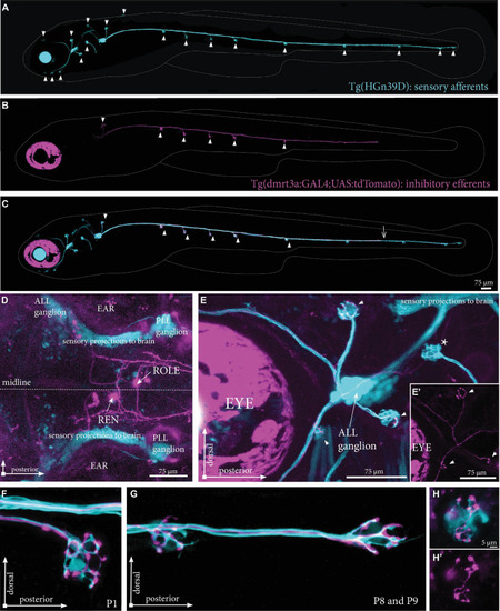
Projections by single inhibitory efferent cells. (A) Overview of the sensory afferent projections in Tg(HGn39D) at 5 dpf. Arrowheads indicate sites of neuromast innervation. (B) A single labeled inhibitory efferent neuron in Tg(dmrt3a:GAL4;UAS:tdTomato) showing partial coverage of the lateral line by its projections. Arrowheads indicate sites of neuromast innervation. (C) Overlay of A and B. Arrowheads indicate sites of afferent and efferent innervation of neuromasts. Arrow indicates the growth cone of the inhibitory efferent projection. Note that A-C do not show the original confocal image, but a modified version to show the lateral line projections only; for the original images, please see Supplementary Figure 2. (D) Top view of a Tg(dmrt3a:GAL4;UAS:tdTomato), where only a single cell in the REN and a single cell in the CEN (ROLE) can be seen. (E) Zoomed image of the ALL ganglion of Tg(HGn39D; dmrt3a:GAL4;UAS:tdTomato) reveal a single projection along the lateral line nerve of the ALL. Arrowheads indicate sites of neuromast innervation by the inhibitory efferent. Asterisks indicate neuromasts without inhibitory efferent innervation [(E’) shows the inhibitory efferent projection only]. (F–G) Higher magnification of Tg(HGn39D; dmrt3a:GAL4;UAS:tdTomato), showing the sensory afferents and a single efferent projection innervating a neuromast at P1 (F) and at P8 and P9 (G). (H) Example of a neuromast that is only partially innervated by the single inhibitory efferent [(H’) shows the inhibitory efferent projection only]. ALL, anterior lateral line; PLL, posterior lateral line; REN, the rostral efferent nucleus; ROLE, the rhombencephalic octavolateral efferent neuron; RELL, the rhombencephalic efferent neuron to the lateral line.
|
