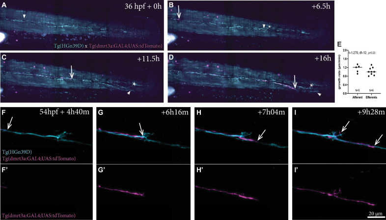
Time-lapse recordings of sensory afferent and inhibitory efferent processes growing along the PLL. (A–D) Time frames of time-lapse imaging made by digital light-sheet microscopy in a Tg(HGn39D; dmrt3a:GAL4;UAS:tdTomato) larva. Arrowheads indicate the growth cone for the sensory afferent projection and arrows indicate the growth cone of the inhibitory efferent projection. Asterisks indicates projections on the other side of the larvae’s body visible in the maximum intensity projections. (E) The growth rate between the sensory afferent projections (1.15 ± 0.18 μm/min) and inhibitory efferent projections (1.0 ± 0.17 μm/min) was not significantly different (t = 1.581, df = 14, p = 0.14). (F–I) Higher magnification time-lapse confocal imaging of the synaptic terminals of afferent processes and a single inhibitory efferent process [(F’–I’) show inhibitory efferent projection only]. Recordings show innervation of a neuromasts by sensory afferents prior to inhibitory efferent innervation. Arrows indicate the growth cone of the inhibitory efferent. Age of larva in (A) 36 hpf; Age of larva in (F) is 54 hpf.
Acknowledgments
This image is the copyrighted work of the attributed author or publisher, and
ZFIN has permission only to display this image to its users.
Additional permissions should be obtained from the applicable author or publisher of the image.
Full text @ Front. Neuroanat.
Your Input Welcome
Thank you for submitting comments. Your input has been emailed to ZFIN curators who may contact you if
additional information is required.
Oops. Something went wrong. Please try again later.