
- Title
-
Use of melatonin as an inhibitor of apoptotic process for cryopreservation of zebrafish (Danio rerio) embryos
- Authors
- Castro, P.L., Ferraz, A.L.J., Patil, J.G., Ribeiro, R.P.
- Source
- Full text @ Braz. J. Biol.
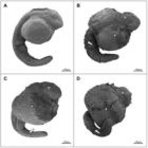
Representative scanning electron micrographs of vitrified and non-vitrified zebrafish (Danio rerio) embryos. (A) Control group; (B-D) Vitrified with 0, 1 µM and 1 mM melatonin respectively. Arrows, arrow head and asterisk point to invaginations and perforations, rupture of the vitelline membrane and wrinkling of the epidermis respectively. |
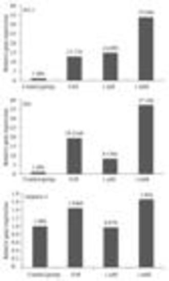
Bax, bcl-2 and caspase-3 mRNA expression levels in zebrafish (Danio rerio) embryos for the control group and vitrified treatments with 0, 1 µM and 1 mM melatonin respectively obtained by the 2-ΔΔCT method. Boxes with same letter are not significantly different from one another (P > 0.05). |
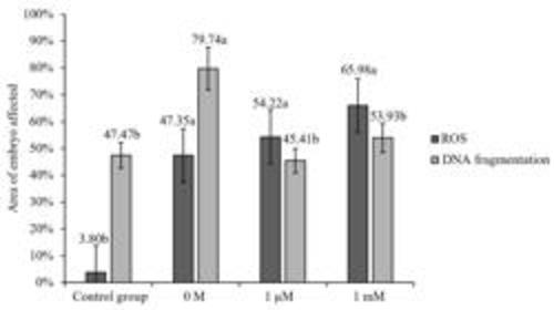
Percentage area of zebrafish (Danio rerio) embryos affected by reactive oxygen species (ROS) formation and DNA fragmentation after vitrification for the control group and vitrified treatments with 0, 1 µM and 1 mM melatonin respectively. Bars with different letter within the same assay were significantly different from one another (P < 0.05). |

Representative images of zebrafish (Danio rerio) embryos showing reactive oxygen species (ROS) formation (A-D) and DNA fragmentation (E-H) following vitrification for the control group and vitrified treatments with 0, 1 µM and 1 mM melatonin respectively. Arrows and arrow heads point to tissues affected in the body axis and those in close proximity to the yolk respectively. |