
Image
|
Figure Caption
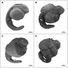
Fig. 1 Representative scanning electron micrographs of vitrified and non-vitrified zebrafish (Danio rerio) embryos. (A) Control group; (B-D) Vitrified with 0, 1 µM and 1 mM melatonin respectively. Arrows, arrow head and asterisk point to invaginations and perforations, rupture of the vitelline membrane and wrinkling of the epidermis respectively.
Acknowledgments
This image is the copyrighted work of the attributed author or publisher, and
ZFIN has permission only to display this image to its users.
Additional permissions should be obtained from the applicable author or publisher of the image.
Full text @ Braz. J. Biol.