Chemical structures of (A) crocetin and (B) crocin.

Crocetin and crocin inhibited SIV formation in Tg(fli1:EGFP) zebrafish embryos. Zebrafish embryos at 24 hpf were treated by (A,A’) 0.1% DMSO (Control) (E,E’) 50 ng/ml VRI, (B-D,B’-D’) crocetin, or (F–I,F’–I’) crocin for 48 h. (A’–I’) Magnified views (100× magnification) of the panels labelled A to I (40× magnification). Mature SIVs are indicated by white arrows. Moderately defective SIVs are indicated with yellow arrows and severely defective SIVs are indicated with red arrows (J and K) Quantification of the total area of SIVs reduced by crocetin and crocin. Data are percentages of the control, measured as means ± SD (10 zebrafish embryos per well from three time-independent experiments, n = 3). Scale bar = 200 μm. Statistical analysis was performed by one-way ANOVA followed by the Dunnett’s test. *p<0.05 and ***p<0.001 versus control group.

Representative HPLC profiles for (A) reference compounds of crocetin and crocin (B) Compounds detected in zebrafish larvae homogenates collected within 72 hpf after crocin treatment (100 μM) (C) Compounds detected in zebrafish larvae homogenates collected within 72 hpf after crocetin treatment (20 μM).

Cytotoxicity of crocetin and crocin on HUVECs (A) HUVECs were treated with different concentrations of crocetin (0.2, 0.3, 0.5, and 1 mM) and (B) crocin (1, 2 and 4 mM) for 24 h and examined by XTT assay. The IC50 value of crocetin (A) is 372.6 μM. Data are means ± SD of three independent experiments (n = 3) as a percentage of control. Statistical analysis was performed by one-way ANOVA followed by Dunnett’s test. **p<0.01; ***p<0.001 versus control group.

Crocetin and crocin inhibited endothelial cell migration and capillary-like tube formation. (A) Wound healing assay for HUVEC migration after 20 h of incubation with vehicle control or drug treatments. (B) The migratory ability was evaluated by measuring the mean length of the scraped area of each well and comparing it to the control group. Yellow dashed lines indicated the wound edges. (C) Morphological features of the capillary-like tube formation of HUVECs in Matrigel after 6 h of incubation with vehicle control or drug treatments. (D) The tube formation ability was evaluated by measuring the total tube length of HUVECs and comparing it to the control group. Scale bar = 200 μm. Data are means ± SD of three independent experiments. Statistical analysis was performed by one-way ANOVA followed by Dunnett's test. *p<0.05 and ***p<0.001 versus control group.

Crocetin and crocin inhibited the activation of VEGFR2 and its downstream signaling pathways. HUVECs were starved for 2 h and then pretreated with crocetin (10, 20 and 40 μM) or crocin (100, 200 and 400 μM) for 4 h before being stimulated by VEGF (50 ng/ml) for 15 min. Western blot assay was used for investigating the expression levels of the major proteins involved in VEGF-mediated angiogenesis signaling in HUVECs. Crocetin and crocin down-regulated the expression levels of (A) p-VEGFR2 (B) p-SRC and p-FAK, and (C) p-MEK and p-ERK. Protein expression levels were quantified by densitometry. Results are percentages relative to control; means ± SD of three independent experiments (n = 3) are shown. Statistical analysis was performed by one-way ANOVA followed by Dunnett’s test. *p<0.05; **p<0.01 and ***p<0.001 versus VEGF treatment group. #p<0.05; ##p<0.01 and ###p<0.001 versus control group.

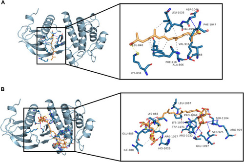
Molecular interactions between crocetin or crocin with VEGFR2. Three-dimensional view of crocetin and crocin located in the binding site of VEGFR2 are shown in (A) and (B), with affinities of −8.6 and −8.4 kcal/mol, respectively. Hydrophobic interactions (dashed lines), hydrogen bonds (blue lines) and important residues of the binding site are shown.

Crocetin and crocin inhibited angiogenesis via the inhibition of VEGFR2 signaling and downstream SRC, FAK, MEK, ERK kinase activation.

Acknowledgments
This image is the copyrighted work of the attributed author or publisher, and ZFIN has permission only to display this image to its users. Additional permissions should be obtained from the applicable author or publisher of the image. Full text @ Front Pharmacol