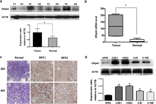
ERp44 was highly expressed in NPC. a Western blot analysis was performed to reveal ERp44 expression in tissues. Tumor: Nasopharyngeal squamous cell carcinoma tissues. Normal: Inflammatory nasopharyngeal epithelium tissues. ACTB was used as a control. The bar demonstrated the expression of ERp44 relative to ACTB by densitometry. b qRT-PCR was used to detect ERp44 expression in NPC tissues and inflammatory nasopharyngeal tissues. c Representative results of immunohistochemical staining. Left: Inflammatory nasopharyngeal epithelium tissues had lower expression of ERp44 (up: × 20) (down: × 40). Middle and Right Line: NPC tissues had higher expression of ERp44 (up: × 20) (down: × 40). d Western blot was used to detect ERp44 expression in NPC cell lines CNE1, CNE2, 5-8F, 6-10B and normal nasopharyngeal epithelial cell NP69. Data represent mean ± SEM. *p < 0.05

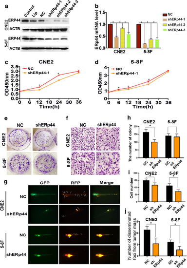
Interference of ERp44 expression could inhibit the malignant phenotype of NPC cells. a CNE2 and 5-8F cells were transfected with shRNA targeting ERp44 (sh-1, sh-2, sh-3) or a scrambled sequence (NC). b qRT-PCR was used to detect the mRNA level of ERp44 after the transfection. c, d CCK8 was used to determine cell proliferation after the treatment of shERp44 in CNE2 and 5-8F. e, h Knockdown of ERp44 in CNE2 could significantly reduce colony formation. We showed the representative images and the quantification analysis. f, i The cells migrated through the membrane in a transwell after ERp44 was knocked down. We showed the representative images of the migrated cells. g, jTg(fli1a: EGFP) transgenic zebrafish was used to detect cell metastasis. Tumor cells were stained in red. The asterisks represented tumor cells in primary sites. The arrows represented tumor cells in disseminated foci. We observed the number of disseminated foci from primary sites after cells were treated with sh-ERp44. All experiments were repeated three times. Data represent mean ± SEM. *p < 0.05

PHENOTYPE:
Fish:
Condition:
Observed In:
Stage: Pec-fin

ERp44 could interact with ACLY in NPC cells. a The results from string protein interaction network, it showed the proper proteins that might interact with ERp44. b Co-IP was used to confirm the interaction of ERp44 with ACLY. c The mutant variants of ERp44 containing the tag HA, mutations in the thioredoxin region (ERp44-Δ 1, mutant in 30–138), mutations in other than thioredoxin region (ERp44-Δ 2, mutant in 139–402). d Co-IP confirmed that ERp44-Δ1was the interaction region with ACLY in 293 T and CNE2 cells. e Western blot showed that when ERp44 was knockdown, there was similar tread of expression change in ACLY. All experiments were repeated three times. Data represent mean ± SEM. *p < 0.05

The binding between ERp44 with ACLY was critical for ERp44-mediated regulation of NPC metastasis. a, b Transwell migration assay showed that downregulating the expression of ACLY could inhibit the migration of NPC cells. The migrated cells were less in ACLY-wt cells co-transfected with ERp44 Δ1 cells compared with that in wild-type cells or Δ2 mutant cells. c, d Wound-healing assay showed cells treated with ERp44 Δ1 migrated slower than other two groups. Representative images of cells migration were shown at 0 and 24 h with a microscope. The relative migrated width was calculated by the wound width/the distance measured at 0 h. The histogram showed the relative distance of wound. e Western blot showed that when ACLY was downregulated, E-cadherin was increased, whereas vimentin was reduced. The histogram showed the expression of E-cadherin or Vimentin relative to ACTB. f Western blot showed that when co-transfected ACLY-wt cells with wild- type ERp44, ERp44 Δ1or ERp44 Δ2 mutant cells, only ERp44 Δ1 could increase the expression of E-cadherin and decrease Vimentin. The histogram showed the expression of proteins relative to ACTB. All experiments were repeated three times. Data represent mean ± SEM. *p < 0.05

ERp44 promoted NPC cells growth in vivo. a Nude mice were subcutaneously injected with CNE2 cells transfected with NC or shERp44. We showed the representative pictures of NPC xenografts in nude mice and tumors. b The volume of tumors treated with shERp44 were smaller than control (n = 5 per group). c Immunohistochemistry was used to access ERp44 and ACLY expression in xenografts, we showed the representative pictures. d The correlation of ERp44 with ACLY expression in xenografts derived from nude mice. e Western blot showed E-cadherin was upregulated in shERp44 tumor sections, while Vimentin was downregulated. Experiments were repeated three times. Data represent mean ± SEM. *p < 0.05

Acknowledgments
This image is the copyrighted work of the attributed author or publisher, and ZFIN has permission only to display this image to its users. Additional permissions should be obtained from the applicable author or publisher of the image. Full text @ J Transl Med