IMAGE

Fig. 1

ID
ZDB-IMAGE-210224-10
Source
Figures for Tian et al., 2021
Image
Figure Caption

Fig. 1

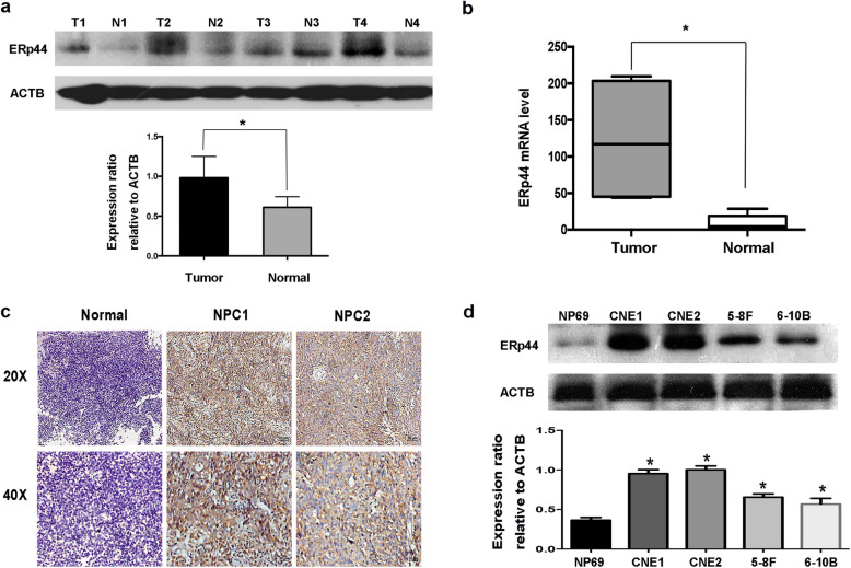
ERp44 was highly expressed in NPC. a Western blot analysis was performed to reveal ERp44 expression in tissues. Tumor: Nasopharyngeal squamous cell carcinoma tissues. Normal: Inflammatory nasopharyngeal epithelium tissues. ACTB was used as a control. The bar demonstrated the expression of ERp44 relative to ACTB by densitometry. b qRT-PCR was used to detect ERp44 expression in NPC tissues and inflammatory nasopharyngeal tissues. c Representative results of immunohistochemical staining. Left: Inflammatory nasopharyngeal epithelium tissues had lower expression of ERp44 (up: × 20) (down: × 40). Middle and Right Line: NPC tissues had higher expression of ERp44 (up: × 20) (down: × 40). d Western blot was used to detect ERp44 expression in NPC cell lines CNE1, CNE2, 5-8F, 6-10B and normal nasopharyngeal epithelial cell NP69. Data represent mean ± SEM. *p < 0.05

Acknowledgments
This image is the copyrighted work of the attributed author or publisher, and ZFIN has permission only to display this image to its users. Additional permissions should be obtained from the applicable author or publisher of the image. Full text @ J Transl Med