FIGURE SUMMARY
Title

Adriamycin does not damage podocytes of zebrafish larvae

Authors
Schindler, M., Blumenthal, A., Moeller, M.J., Endlich, K., Endlich, N.
Source
Full text @ PLoS One

Evaluation of phenotype, autofluorescence, vascular eGFP fluorescence, survival and edema rate of larvae after treatment.

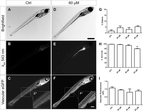
Treated larvae did not develop periocular or pericardial edemas at a significant rate (A, D, G). However, high concentrations of ADR caused an increase in mortality of the larvae (H). All doses of ADR led to a strong accumulation of ADR in the intestinal tract which caused a remarkable autofluorescence over a wide spectrum (B, E). Quantification of the vascular eGFP fluorescence of DBP larvae (Tg(-3.5fabp10a:gc-eGFP) after ADR treatment did not reveal a significant vascular reduction of the eGFP-DBP fusion protein (~68 kDa) (C, F, I). Scale bar in D represents to 200 μm, in F* to 50 μm. *P = 0,03.

Glomeruli of ADR-treated larvae did not show morphological alterations demonstrated by HE staining on histological sections.

A loss of podocytes or a Bowman‘s space edema could not be observed (A-D). n: notochord, da: dorsal aorta, c: capillary, p:podocyte, bs: Bowman‘s space. Scale bar represents 10 μm.

Evaluation of wt1a expression <italic>in vivo</italic> and immunostaining of cryosections for the slit membrane protein nephrin in ADR-treated larvae.

2-PM in vivo imaging revealed no morphological changes or glomerular abnormalities in treated larvae (A). Immunostainings showed that ADR treatment neither reduces the expression of the pronephric transcription factor wt1a nor changes the expression pattern of nephrin (B). RT-PCR (C) and RT-qPCR (D) confirmed that nephrin mRNA expression was not altered in ADR-treated larvae at 9 dpf. Scale bars represent 10 μm.

EXPRESSION / LABELING:
Genes:
Fish:
Condition:
Anatomical Term:
Stage: Days 7-13

Morphology and ultrastructure of larval glomeruli.

Morphological investigations by Richardson‘s staining of semithin sections (500 nm) did not show abnormalities of the glomerular morphology due to ADR treatment (A, B). Ultrastructural analysis of the pronephric filtration barrier confirmed the abscence of a glomerular phenotype (C-F). The interdigitating pattern of podocyte foot processes (asterisk in E and F) is present in both control was well as 60 μM ADR-treated larvae. The slit membrane as a highly organized structure connecting foot processes was not affected by ADR (arrows in E and F). Scale bars in A and B represent 10 μm, in C and D 1 μm, in E and F 200 nm.

Acknowledgments
This image is the copyrighted work of the attributed author or publisher, and ZFIN has permission only to display this image to its users. Additional permissions should be obtained from the applicable author or publisher of the image. Full text @ PLoS One