
- Title
-
p53 isoform Δ113p53 promotes zebrafish heart regeneration by maintaining redox homeostasis
- Authors
- Ye, S., Zhao, T., Zhang, W., Tang, Z., Gao, C., Ma, Z., Xiong, J.W., Peng, J., Tan, W.Q., Chen, J.
- Source
- Full text @ Cell Death Dis.

|
|
|
|

( |
|
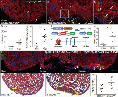
Δ113p53:GFP was not detectable in the surgery hearts of p53M214K mutant fish at 14 dpa. (A-B) Cryosections of Tg(Δ113p53:GFP) hearts of p53+/+ (A) and p53M214K mutant (B) at 14 dpa were immunostained by anti-GFP (in green) and anti-MHC (MF20) (in red) antibodies. The nucleus were stained with DAPI (in blue). The representative picture was taken from 3 hearts in each group. The white arrow heads indicate wounding site. Scale bar, 50 μm. |
|
Validation of Tg(Δ113p53:CreER; β-act2:RSG) zebrafish. (A) Western blot with a monoclonal antibody against zebrafish Δ113p53 was performed to analyze the induction of Δ113p53 upon the treatment of a DNA damage drug, camptothecin (Campt). The Tg(Δ113p53:CreER; β-act2:RSG) zebrafish embryos at 1 day post fertilization (dpf) were treated with Campt for 24 hours. Afterwards, a part of untreated and Campt-treated embryos were divided and treated with 4HT for 2 hours. β-actin was used as the protein loading control. (B-E) Life images of red (DsRed) (B, C, D, E), green (EGFP) (B’, C’, D’, E’) and bright field (B”, C”, D”, E”) in Tg(Δ113p53:CreER; β-act2:RSG) zebrafish embryos treated with either Campt (C, C’, C”), or 4HT (D, D’, D”), or both (E, E’, E”) as described above. Scale bar, 500 μm. |
|
Δ113p53M/M mutant and WT uninjured hearts are comparative. (A-B) Trichrome Masson’s staining on the crysections of uninjured Δ113p53+/+ (A) and Δ113p53M/M mutant hearts (B) at 9 months of age. The representative picture was taken from 5 hearts in each group. Scale bar, 50 μm. |
|
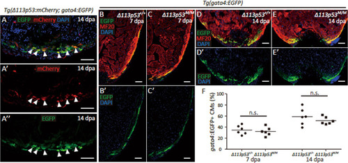
style='font-size:15px;line-height: 29.333335876464844px;font-family: "Times New Roman";'>Validation of Tg(Δ113p53:mCherry) zebrafish. (A) Diagram showing the Tg(Δ113p53:mCherry) reporter driven by Δ113p53promoter. Δ113p53-P (blue arrow): the 3.6 kb DNA fragment from the upstream of Δ113p53 transcription start site; mCherry (red bar): the coding region of mCherry. (B-C) Live images of red (mCherry) fluorescence in Tg(Δ113p53:mCherry) zebrafish embryos treated with Campt (C) or without Campt (B) for 24 hours. Scale bar, 500 μm. |
|
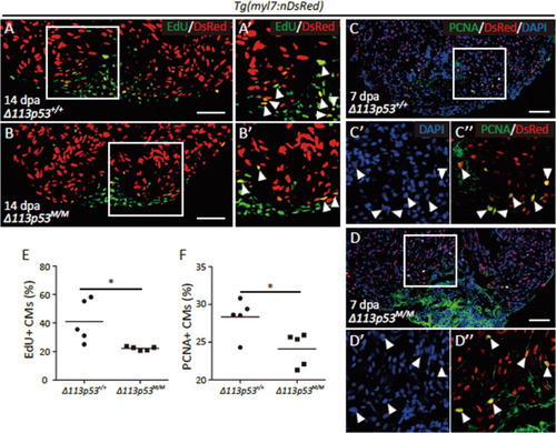
Depletion of Δ113p53 has little effects on DNA damage response and apoptotic activity during heart regeneration. (A-B) Cryosections of Tg(myl7:nDsRed); Δ113p53+/+ (A) and Tg(myl7:nDsRed); Δ113p53M/M (B) hearts at 14 dpa were co-stained by anti-DsRed (in red) and anti-γ-H2AX (in green) antibodies. Framed areas were magnified in A’ and B’. (C-D) TUNEL assay (in green) and co-immunostaining with anti-DsRed antibody (in red) were performed on cryosections of Tg(myl7:nDsRed);Δ113p53+/+ (C) and Tg(myl7:nDsRed); Δ113p53M/M (D) hearts at 14 dpa. Framed areas were magnified in C’ and D’. The representative picture was taken from 6 hearts in each group. Scale bar, 50 μm. |