FIGURE SUMMARY
Title

Cytotoxic Evaluation and Anti-Angiogenic Effects of Two Furano-Sesquiterpenoids from Commiphora myrrh Resin

Authors
S Alqahtani, A., Nasr, F.A., Noman, O.M., Farooq, M., Alhawassi, T., Qamar, W., El-Gamal, A.
Source
Full text @ Molecules

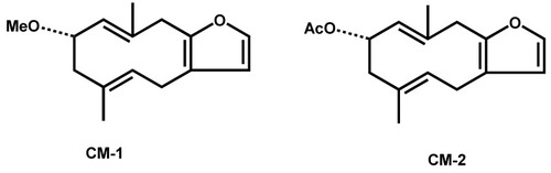
Chemical structures of 2-methoxyfuranodiene (left) and 2-acetoxyfuranodiene (right) isolated from the chloroform fraction of C. myrrh.

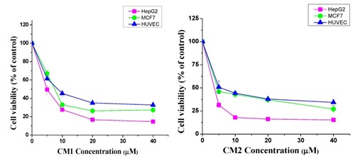
Cytotoxic effects of compounds CM1 and CM2 on different human cell lines. The viability was measured by an MTT assay. Cells were cultured as described in the methods and treated with various concentrations of CM1 and CM2 (1–40 µM) for 48 h. The Student’s t-test was used to calculate the statistical differences. The data are presented as the mean ± S.D. (* p < 0.05, ** p < 0.01, and *** p < 0.001 were considered significant compared to the control) for the three independent experiments carried out in triplicate.

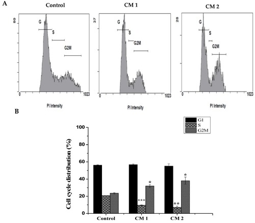
Compounds CM1 and CM2 induced cell cycle arrest. HepG2 cells were exposed to the IC50 for 48 h and analysis was determined as described. (A) Distribution of cell phases after treatment with compounds CM1 and CM2. (B) Quantitative analysis of data obtained. Data represented as the mean ± SD (n = 3). (* p < 0.05, ** p < 0.01, *** p < 0.001 vs. control)

HepG2 cell death after treatment with compounds CM1 and CM2. Cells were exposed to the IC50 for 48 h, and cells undergoing either apoptosis or necrosis were assessed using co-staining with Annexin-V/FITC-PI dyes. (A) Representative scatter plots of PI (y-axis) vs. FITC-Annexin V (x-axis); live cells (A3) were negative for both dyes, (A4) early apoptotic cells were positive for only FITC -Annexin V, (A2) late apoptotic cells were stained with both dyes and(A1) Necrotic cells were stained with PI only. (B) Percentage of viable, early apoptotic, and late apoptotic + necrotic cells. Data are presented as the means ± SD of the triplicate experiments. ** p < 0.05 and *** p < 0.01 vs. control.

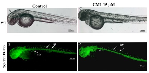
2-methoxyfuranodiene inhibited the secondary angiogenic blood vessel formation in zebrafish embryos. (A) Bright field image of a Tg(fli1:EGFP) embryo that was exposed to 0.1% methanol as a control. The embryos developed normally. (B) The fluorescent image of a Tg (fli1: EGFP) embryo at 72 hpf. The green color indicates all the blood vessels. The white arrows show the inter-segmental blood vessels in the trunk of the mock-treated zebrafish embryo, which were the first angiogenic blood vessels that formed at around 48 hpf. The emergence of the second angiogenic blood vessels can be seen on the trunk of the control embryo. (C) The bright field image of a Tg (fli1: EGFP) embryo treated with 10 µM of CM1. The bright field image shows the normal development of the embryos. (D) The fluoresce image of a Tg (fli1: EGFP) zebrafish embryo treated with 15 µM of CM1 showed the normal formation of the inter-segmental blood vessels. However, the treated embryos do not have the sub-intestinal vein blood vessels (indicated by white asterisks).

2-acetoxyfuranodiene decreased the formation of angiogenic blood vessels during zebrafish embryonic development. (A) Bright field image of a Tg (fli1: EGFP) embryo that was exposed to 0.1% methanol as a control. The embryos developed normally. (B) The fluorescent image of a Tg (fli1: EGFP) embryo at 72 hpf. The green color indicates all the blood vessels. The white arrows show the inter-segmental blood vessels in the trunk of the control embryo, which were the first angiogenic blood vessels that formed at around 48 hpf. The emergence of the second angiogenic blood vessels can be seen on the trunk of the control embryo. (C) A Tg (fli1: EGFP) embryo treated with 15 µM of CM2. The bright field images show that CM2 at 15 µM affected the normal development of zebrafish embryos. The embryos were much smaller in size, the body was curved, and there was no pigmentation. (D) The CM2 treatment disrupted the inter-somatic blood vessel formation in the treated zebrafish embryos, the majority of the inter-somatic blood vessels did not form (white asterisks), and some blood vessels formed but they did not grow and failed to connect to dorsal longitudinal anastomotic vessel. Similarly, the sub-intestinal vein did not form in the transgenic zebrafish embryos that were treated with CM2.

Acknowledgments
This image is the copyrighted work of the attributed author or publisher, and ZFIN has permission only to display this image to its users. Additional permissions should be obtained from the applicable author or publisher of the image. Full text @ Molecules