FIGURE SUMMARY
Title

Bucillamine Prevents Afatinib-Mediated Inhibition of Epidermal Growth Factor Receptor Signaling

Authors
Nishiya, N., Murai, M., Hosoda, A., Yonezawa, H., Omori, N.
Source
Full text @ Pharmaceuticals (Basel)

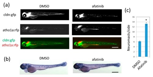
Afatinib induces the development of extra lateral line neuromasts in zebrafish larvae. (a,b) Neuromasts of 72 hpf larvae treated with DMSO (left) or afatinib (right) from 26 hpf were detected by fluorescence from cldn:gfp; atoh1:rfp transgenic larvae (a) or by endogenous ALP activity (b). (a) Fluorescence from cldnb:gfp highlighted neuromast cells and the epidermis, and fluorescence from atoh1a:rfp highlighted neuromast hair cells. GFP and RFP images were merged. Compared to DMSO treatment, increased neuromasts were observed in afatinib-treated zebrafish larvae. Scale bar = 500 µm. (c) The number of neuromasts on one side were counted. The graph indicates mean ± SEM (n = at least 54). * p < 0.01; hpf, hours postfertilization; DMSO, dimethyl sulfoxide; ALP, alkaline phosphatase; GFP, green fluorescent protein; RFP, red fluorescent protein; SEM, standard error of the mean.

Bucillamine, an antirheumatic drug, suppressed afatinib-induced extra lateral line neuromast development. (a) Structures of the test compound, acrolein, and MESNA are shown. (b) Clinically available sulfhydryl compounds were evaluated for afatinib quenchers. Neuromasts of 72 hpf larvae treated with the indicated test compound were detected by fluorescence of cldnb:gfp. Scale bar = 500 µm. (c) The number of neuromasts on one side were counted. The graph indicates the mean ± SD (n = at least 4). * p < 0.01; MESNA, sodium 2-mercaptoethanesulfonate; hpf, hours postfertilization; SD, standard deviation.

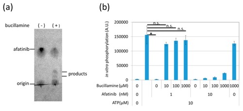
Bucillamine inhibited afatinib-mediated EGFR kinase inhibition in vitro. (a) 10 μM afatinib was incubated with DMSO or 1 mM bucillamine for 90 min at 25 °C. The reaction products were analyzed by TLC with UV detection. (b) A recombinant EGFR kinase domain was pre-incubated with 1 or 10 nM afatinib in the presence or absence of 10–1000 μM bucillamine. Then, 10 μM ATP was added to the kinase in order to initiate phosphorylation of an artificial substrate peptide. Phosphorylation levels were measured by ELISA using antiphosphotyrosine antibody. The graph indicates the mean ± SEM (n = at least 3). DMSO, dimethyl sulfoxide; EGFR, epidermal growth factor receptor; TLC, thin-layer chromatography; UV, ultraviolet; ATP, adenosine triphosphate; ELISA, enzyme-linked immunosorbent assay; SEM, standard error of the mean.

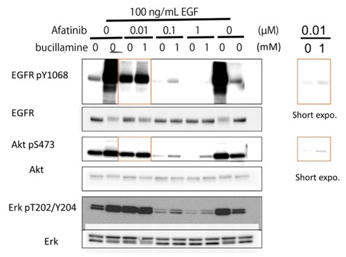
Bucillamine inhibited afatinib-mediated inhibition of EGFR autophosphorylation and downstream signaling. A431 cells expressing endogenous WT EGFR were pre-incubated with 0.01–1 μM afatinib in the presence or absence of 1 mM bucillamine. Then, 100 ng/mL of EGF was added to the culture medium. After 15 min incubation, cell lysates were prepared and analyzed by Western blotting with specific antibodies, as indicated. Shorter-exposure images are shown on the right. EGFR, epidermal growth factor receptor; WT, wild type.

Acknowledgments
This image is the copyrighted work of the attributed author or publisher, and ZFIN has permission only to display this image to its users. Additional permissions should be obtained from the applicable author or publisher of the image. Full text @ Pharmaceuticals (Basel)